
What are Feline Injection-Site Sarcomas (FISS)?
Maigan Espinili Maruquin
I. Characteristics / Epidemiology
The feline injection-site sarcomas (FISS) were first reported on 1991 (Hendrick and Goldschmidt 1991). With the implementation of stricter vaccination and development of vaccines for rabies and FeLV, the increased incidence of vaccine reactions was recognized (Hendrick and Dunagan 1991, Kass, Barnes et al. 1993, Hartmann, Day et al. 2015, Saba 2017). With this, recommendations were to use the term ‘vaccine-associated sarcomas’, however, studies show that aside from vaccines are other non-vaccinal injectables in the subcutis or muscle can also cause chronic inflammatory response which led to reclassification as ‘feline injection-site sarcomas’ (FiSSs) (Martano, Morello et al. 2011, Hartmann, Day et al. 2015).
The FISS develops in 1–10 of every 10,000 vaccinated cats wherein malignant skin tumors of mesenchymal origin develops (Zabielska-Koczywąs, Wojtalewicz et al. 2017). It has been described as secondary to inflammation in different organs like eye (PEIFFER, MONTICELLO et al. 1988), uterus (Jelínek 2003) and muscle or skin after placement of non-absorbable suture or microchips (Buracco, Martano et al. 2002) (Bowlt 2015). Between three months to 10 years after vaccination, the development of FISS can occur (Hendrick, Shofer et al. 1994, McEntee and Page 2001) (Esplin, D. G., et al., 1993). Whereas, a study reported that the younger cats developed tumor at the vaccination site as compared to the older ones with similar tumors in other body areas with bimodal distribution of age with a peak at 6–7 years and a second at 10–11 years (Kass, Barnes et al. 1993, Martano, Morello et al. 2011).
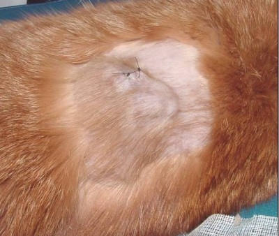
Fig. 01. Saba, C. F. 2017 shows the occurrence of FISS.
(https://doi.org/10.2147/VMRR.S116556)
II. Pathogenesis / Clinical Signs
After investigations, the hypothesis suggests that secondary to chronic and inflammatory response to vaccine or injection, having ultimate malignant transformation of surrounding fibroblasts and myofibroblasts triggers the tumors (Hendrick and Brooks 1994, Hartmann, Day et al. 2015, Saba 2017)( Hendrick MJ., 1999).
Fig. 02. (Cecco, B.S., et al., 2019) The sites where FISS occurs
(https://doi.org/10.1016/j.jcpa.2019.08.009)
Reports show significant correlation between the rabies and/ or FeLV vaccinations in the development of FISS (Hendrick, Goldschmidt et al. 1992, Kass, Barnes et al. 1993, Hendrick, Shofer et al. 1994). Despite many causes are associated with what triggers the tumor, higher risks are seemed to be coming from vaccines, specifically adjuvanted (Hartmann, Day et al. 2015). Discovered were traces of adjuvants in the inflammatory reaction and later in histological sections (Hendrick and Brooks 1994, Hartmann, Day et al. 2015).
Particles of grey- brown material in the necrotic centre and within the cytoplasm of macrophages were reported consistent with an inflammatory reaction (Hendrick and Dunagan 1991, Hendrick and Brooks 1994, Martano, Morello et al. 2011). The infiltrates reported includes macrophages often having cytoplasmic material, giant cells, lymphocytes and mixed neutrophils and eosinophils. Further, identified in the tumors were cytokines, growth factors and mutations in tumor suppressor genes (Ladlow 2013, Carneiro, de Queiroz et al. 2018).
While fibrosarcoma is commonly diagnosed, some histological types were also reported to include: malignant fibrous histiocytoma, rhabdomyosarcoma, myxosarcoma, liposarcoma, nerve sheath tumor, poorly differentiated sarcomas, and extraskeletal osteosarcoma and chondrosarcoma (Esplin, McGill et al. 1993, Hendrick and Brooks 1994, Hershey, Sorenmo et al. 2000, Dillon, Mauldin et al. 2005, Saba 2017). According to Saba. C, 2017, any sarcoma that develops within the vicinity of vaccination or injection site should be considered an FISS and thus, should be treated aggressively.
While tumors are invasive and variable in size, Martano M.E., et al, 2011 reported that large sized may be due to rapid growth. On the other hand, there could also be delayed in appearance due to its interscapular or deep location (Bowlt 2015). The mass can also be mobile or intensely adherent to the underlying tissue which is usually not painful, but solid and may be cystic (Bowlt 2015). These tumors that develop commonly in sites of injection can reach several centimetres in diameter within a few weeks (Martano, Morello et al. 2011).
Since not all cats develop this tumor after vaccination, suggestions are due to genetic predisposition, with higher case of FISS occurrence in siblings of affected cats. Further, some cats may develop more than one FiSS (Hartmann, Day et al. 2015).
III. Staging / Diagnosis
To properly react with the tumor, proper staging shall be performed. Once a histological diagnosis has been confirmed (Bowlt 2015), it requires complete blood count, a serum biochemical panel, urinalysis, 3-view thoracic radiography, lymph node examination by palpation, and ultrasonography of the abdominal cavity and cytology when applicable (Séguin 2002, Zabielska-Koczywąs, Wojtalewicz et al. 2017). Abdominal ultrasound may be required, depending on the location of the tumor. Computed tomography (CT) or magnetic resonance imaging (MRI) of the lesion and the thorax is required to see the actual size and evaluate the extent of the tumor (Cronin, Page et al. 1998, McEntee and Page 2001, Martano, Morello et al. 2011, Rousset, Holmes et al. 2013, Travetti, di Giancamillo et al. 2013, Saba 2017, Zabielska-Koczywąs, Wojtalewicz et al. 2017). Thoracic radiography is then performed to exclude metastatic deseases, which has 10- 24% chances (Saba 2017, Zabielska-Koczywąs, Wojtalewicz et al. 2017). Whereas, there is as high as 45% for the recurrence rate even after performing surgical excision (Cronin, Page et al. 1998)
IV. Treatment
Considering the possibility of misdiagnosing the tumor as a granuloma from small tissue samples, and the fact that these can be heterogeneous, incisional biopsy can be done at sites that can be easily excised (Martano, Morello et al. 2011). The indications for a biopsy are based in 3-2-1 rule (Vaccine-Associated Feline Sarcoma Task Force, 2005; Vaccine-Associated Feline Sarcoma Task Force guidelines, 1999; (Morrison and Starr 2001). This incisional biopsy is strongly recommended for masses that has persisted for >3 months, is >2 cm, and/or is growing over the course of 1 month post injection in the site (Saba 2017).
Radical surgery or wide excision may be recommended. Surgery will be performed with at least 3 cm margins peripherally and one fascial plane deep into the tumor wherein completeness of surgical margins is then considered the most important predictive factor for the FISS treatment. (Zabielska-Koczywąs, Wojtalewicz et al. 2017).
Radiotherapy is recommended often due to the high urate of recurrence, as high as 70% if the radical surgery is insufficient, and to have a disease- free survival (Hendrick and Brooks 1994). When the radiotherapy was performed before the surgery, metastatic seeding during surgery is less likely (Zabielska-Koczywąs, Wojtalewicz et al. 2017). Having both the radical surgery and radiotherapy might be the most efficient way to treat FISS but short- and long-term side effects shall be considered, too (Zabielska-Koczywąs, Wojtalewicz et al. 2017). The side effects of the radiation can be mild and primarily include dry desquamation of the skin, however, the healing of the wound may also be delayed (Saba 2017).
Having chemotherapy is not used as monotherapy, but may serve as neoadjuvant setting in a multimodal approach and cytostatic drugs were used in FiSS treatment (Zabielska-Koczywąs, Wojtalewicz et al. 2017). Further, additional immunotherapy has also promising reults (Jahnke, Hirschberger et al. 2007, Hüttinger, Hirschberger et al. 2008, Hartmann, Day et al. 2015, Jas, Soyer et al. 2015). Studies of cytokine gene transfer techniques as adjuvant-immunological treatment has showed less recurrence rates (Hartmann, Day et al. 2015).
For patients that has FISS, the age, sex, breed, of the FeLV status don’t seem to affect the survival time (Bowlt 2015). After the surgery, recurrence of tumor or metastasis significantly affects survival (Phelps, Kuntz et al. 2011, Bowlt 2015).
V. Prevention
To prevent the occurrence of the FISS, few things must be considered. The choice of injection site should be assessed. It is recommended to have the site recommended for monitoring (Hartmann, Day et al. 2015).
The inflammatory reactions shall also be avoided in the injection sites. It is recommended to give subcutaneous injections as few as possible. Intramuscular tumours develop with a similar frequency, but are more difficult to detect early, thus must also be avoided. Oral drugs or intravenous must be given if needed. In addition to, long-acting irritating substances of injections must be avoided. And finally, cats should be vaccinated no more than necessary (Hartmann, Day et al. 2015).
References
- Cecco, B.S., Henker, L.C., De Lorenzo, C., Schwertz, C.I., Bianchi, R.M., da Costa, F.V.A., Driemeier, D., Pavarini, S.P., Sonne, L. (2019). Epidemiological and Pathological Characterization of Feline Injection Site Sarcomas in Southern Brazil. Journal of Comparative Pathology. 172: 31-36.
- Hendrick MJ. Feline vaccine-associated sarcomas. Cancer Invest. 1999;17(4):273–277
- Esplin , D. G., McGill , L. D., Meninger , A. C. & Wilson , S. R. (1993) Postvaccination sarcomas in cats. Journal of the American Veterinary Medical Association 202, 193-196
- Vaccine-Associated Feline Sarcoma Task Force guidelines. Diagnosis and treatment of suspected sarcomas. J Am Vet Med Assoc. 1999;214(12):1745.
- Vaccine-Associated Feline Sarcoma Task Force. The current understanding and management of vaccine-associated sarcomas in cats. J Am Vet Med Assoc. 2005;226(11):1821–1842.
- Bowlt, K. (2015). “Feline injection site-associated sarcomas.” In Practice 37(1): 2-8.
- Buracco, P., M. Martano, E. Morello and A. Ratto (2002). “Vaccine-associated-like Fibrosarcoma at the Site of a Deep Nonabsorbable Suture in a Cat.” The Veterinary Journal 163(1): 105-107.
- Carneiro, C. S., G. F. de Queiroz, A. C. Pinto, M. L. Z. Dagli and J. M. Matera (2018). “Feline injection site sarcoma: immunohistochemical characteristics.” Journal of Feline Medicine and Surgery 21(4): 314-321.
- Cronin, K., R. L. Page, G. Spodnick, R. Dodge, E. N. Hardie, G. S. Price, D. Ruslander and D. E. Thrall (1998). “Radiation therapy and surgery for fibrosarcoma in 33 cats.” Vet Radiol Ultrasound 39(1): 51-56.
- Dillon, C. J., G. N. Mauldin and K. E. Baer (2005). “Outcome following surgical removal of nonvisceral soft tissue sarcomas in cats: 42 cases (1992-2000).” J Am Vet Med Assoc 227(12): 1955-1957.
- Esplin, D. G., L. D. McGill, A. C. Meininger and S. R. Wilson (1993). “Postvaccination sarcomas in cats.” J Am Vet Med Assoc 202(8): 1245-1247.
- Hartmann, K., M. J. Day, E. Thiry, A. Lloret, T. Frymus, D. Addie, C. Boucraut-Baralon, H. Egberink, T. Gruffydd-Jones, M. C. Horzinek, M. J. Hosie, H. Lutz, F. Marsilio, M. G. Pennisi, A. D. Radford, U. Truyen and K. Möstl (2015). “Feline injection-site sarcoma: ABCD guidelines on prevention and management.” Journal of Feline Medicine and Surgery 17(7): 606-613.
- Hendrick, M. J. and J. J. Brooks (1994). “Postvaccinal sarcomas in the cat: histology and immunohistochemistry.” Vet Pathol 31(1): 126-129.
- Hendrick, M. J. and C. A. Dunagan (1991). “Focal necrotizing granulomatous panniculitis associated with subcutaneous injection of rabies vaccine in cats and dogs: 10 cases (1988-1989).” J Am Vet Med Assoc 198(2): 304-305.
- Hendrick, M. J. and M. H. Goldschmidt (1991). “Do injection site reactions induce fibrosarcomas in cats?” J Am Vet Med Assoc 199(8): 968.
- Hendrick, M. J., M. H. Goldschmidt, F. S. Shofer, Y. Y. Wang and A. P. Somlyo (1992). “Postvaccinal sarcomas in the cat: epidemiology and electron probe microanalytical identification of aluminum.” Cancer Res 52(19): 5391-5394.
- Hendrick, M. J., F. S. Shofer, M. H. Goldschmidt, J. C. Haviland, S. H. Schelling, S. J. Engler and J. M. Gliatto (1994). “Comparison of fibrosarcomas that developed at vaccination sites and at nonvaccination sites in cats: 239 cases (1991-1992).” Journal of the American Veterinary Medical Association 205(10): 1425-1429.
- Hershey, A. E., K. U. Sorenmo, M. J. Hendrick, F. S. Shofer and D. M. Vail (2000). “Prognosis for presumed feline vaccine-associated sarcoma after excision: 61 cases (1986-1996).” J Am Vet Med Assoc 216(1): 58-61.
- Hüttinger, C., J. Hirschberger, A. Jahnke, R. Koestlin, T. Brill, C. Plank, H. Küchenhoff, S. Krieger and U. Schillinger (2008). “Neoadjuvant gene delivery of feline granulocyte-macrophage colony-stimulating factor using magnetofection for the treatment of feline fibrosarcomas: A phase I trial.” The journal of gene medicine 10: 655-667.
- Jahnke, A., J. Hirschberger, C. Fischer, T. Brill, R. Köstlin, C. Plank, H. Küchenhoff, S. Krieger, K. Kamenica and U. Schillinger (2007). “Intra-tumoral Gene Delivery of feIL-2, feIFN-γ and feGM-CSF using Magnetofection as a Neoadjuvant Treatment Option for Feline Fibrosarcomas: A Phase-I Study.” Journal of Veterinary Medicine Series A 54(10): 599-606.
- Jas, D., C. Soyer, P. De Fornel-Thibaud, F. Oberli, D. Vernes, P. M. Guigal, H. Poulet and P. Devauchelle (2015). “Adjuvant immunotherapy of feline injection-site sarcomas with the recombinant canarypox virus expressing feline interleukine-2 evaluated in a controlled monocentric clinical trial when used in association with surgery and brachytherapy.” Trials in Vaccinology 4: 1-8.
- Jelínek, F. (2003). “Postinflammatory sarcoma in cats.” Exp Toxicol Pathol 55(2-3): 167-172.
- Kass, P. H., W. G. Barnes, Jr., W. L. Spangler, B. B. Chomel and M. R. Culbertson (1993). “Epidemiologic evidence for a causal relation between vaccination and fibrosarcoma tumorigenesis in cats.” J Am Vet Med Assoc 203(3): 396-405.
- Ladlow, J. (2013). “Injection site-associated sarcoma in the cat: treatment recommendations and results to date.” J Feline Med Surg 15(5): 409-418.
- Martano, M., E. Morello and P. Buracco (2011). “Feline injection-site sarcoma: Past, present and future perspectives.” The Veterinary Journal 188(2): 136-141.
- McEntee, M. C. and R. L. Page (2001). “Feline vaccine-associated sarcomas.” J Vet Intern Med 15(3): 176-182.
- Morrison, W. B. and R. M. Starr (2001). “Vaccine-associated feline sarcomas.” J Am Vet Med Assoc 218(5): 697-702.
- PEIFFER, R. L., T. MONTICELLO and T. W. BOULDIN (1988). “Primary ocular sarcomas in the cat.” Journal of Small Animal Practice 29(2): 105-116.
- Phelps, H. A., C. A. Kuntz, R. J. Milner, B. E. Powers and N. J. Bacon (2011). “Radical excision with five-centimeter margins for treatment of feline injection-site sarcomas: 91 cases (1998-2002).” J Am Vet Med Assoc 239(1): 97-106.
- Rousset, N., M. A. Holmes, A. Caine, J. Dobson and M. E. Herrtage (2013). “Clinical and low-field MRI characteristics of injection site sarcoma in 19 cats.” Vet Radiol Ultrasound 54(6): 623-629.
- Saba, C. F. (2017). “Vaccine-associated feline sarcoma: current perspectives.” Veterinary medicine (Auckland, N.Z.) 8: 13-20.
- Séguin, B. (2002). “Feline injection site sarcomas.” Vet Clin North Am Small Anim Pract 32(4): 983-995, viii.
- Travetti, O., M. di Giancamillo, D. Stefanello, R. Ferrari, C. Giudice, V. Grieco and J. H. Saunders (2013). “Computed tomography characteristics of fibrosarcoma — a histological subtype of feline injection-site sarcoma.” J Feline Med Surg 15(6): 488-493.
- Zabielska-Koczywąs, K., A. Wojtalewicz and R. Lechowski (2017). “Current knowledge on feline injection-site sarcoma treatment.” Acta Veterinaria Scandinavica 59(1): 47.