
Factor VII Deficiency in Dogs
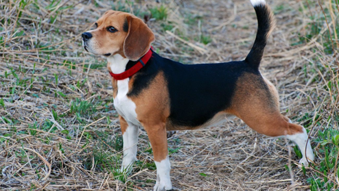
Bioguard Corporation Canine Factor VII (FVII) deficiency is an autosomal recessive genetic disorder that leads to a mild to moderate blood clotting problem in affected dogs. Puppies with the condition may exhibit symptoms such as nosebleeds (epistaxis) and gum bleeding, while adult dogs are more prone to bruising and skin issues like dermatitis. Though bleeding episodes tend to decrease in severity as the dog matures, the disorder still causes ongoing issues throughout the animal’s life. FVII deficiency was first identified in the Migluo breed. Most dogs are diagnosed when they visit a veterinarian due to accidental injury, spontaneous bleeding, genetic screening, or blood tests. Pathogenesis Hemostasis is achieved through a series of events known as the coagulation cascade, which involves two interconnected pathways: the intrinsic and extrinsic pathways. The intrinsic pathway is triggered by spontaneous internal damage to the blood vessel lining, while the extrinsic pathway is activated in response to external trauma. Factor VII (FVII) is a vitamin K-dependent glycoprotein produced in the liver and released into the bloodstream as a single-chain zymogen. Once activated, FVII plays a crucial role in initiating coagulation. Following vascular injury, FVII, along with tissue factor (TF) and calcium, activates factors IX and X, leading to thrombin production. A deficiency in FVII impairs blood clotting, resulting in excessive bleeding during injuries or surgeries. Diagnosis The Buccal Mucosal Bleeding Time (BMBT) is the most commonly used test for measuring bleeding time in small animals. To perform the BMBT, the upper lip is folded back and secured with a gauze strip around the maxilla or both the maxilla and mandible. A small incision is made in the mucosa above the premolars, avoiding areas with visibly engorged vessels. Blood from the incision is gently blotted using filter paper placed near the incision without touching it. A stopwatch starts when the incision is made and stops when no blood crescent forms on the filter paper. To reduce variability, the same person should perform the BMBT whenever possible. Activated Partial Thromboplastin Time (APTT) measures the overall speed of blood clot formation through the intrinsic and common coagulation pathways. It assesses the activity of factors XII, XI, IX, VIII, X, V, II, I, as well as prekallikrein (PK) and high molecular weight kininogen (HK). Prothrombin Time (PT) is used to evaluate the extrinsic and common pathways of coagulation by measuring factors VII, X, V, II, and I. Factor VII deficiency is suspected when a dog presents with a prolonged PT and normal BMBT and APTT. Genetic testing can also identify affected dogs or carriers, especially in certain breeds. Breeds at Risk Canine Factor VII deficiency has been documented in several breeds, including the Beagle, Airedale, Alaskan Klee Kai, American Foxhound, Finnish Hound, German Wirehaired Pointer, Giant Schnauzer, Irish Water Spaniel, Japanese Spitz, Miniature Schnauzer, Papillon/Phalene, Sealyham Terrier, Scottish Deerhound, and Welsh Springer Spaniel. Management There is currently no cure for Factor VII deficiency. However, clinical symptoms can be managed through transfusions with fresh plasma or blood, or by administering recombinant activated human FVII. These treatments provide only temporary relief. Fortunately, dogs with mild to moderate FVII deficiency typically lead normal lives. References Callan MB, Aljamali MN, Margaritis P, Griot-Wenk ME, Pollak ES, Werner P, Giger U, High KA. A novel missense mutation responsible for factor VII deficiency in research Beagle colonies. J Thromb Haemost. 2006 Dec; 4(12):2616-22. Carlstrom LP, Jens JK, Dobyns ME, Passage M, Dickson PI, Ellinwood NM. Inadvertent propagation of factor VII deficiency in a canine mucopolysaccharidosis type I research breeding colony. Comp Med. 2009 Aug;59(4):378-82. Donner J, Kaukonen M, Anderson H, Moller F, Kyostila K, Sankari S, Hytonen M, Giger U, Lohi H. Genetic Panel Screening of Nearly 100 Mutations Reveals New Insights into the Breed Distribution of Risk Variants for Canine Hereditary Disorders. PLoS One. 2016 Aug 15;11(8):e0161005. Kaae JA, Callan MB, Brooks MB. Hereditary factor VII deficiency in the Alaskan Klee Kai dog. J Vet Intern Med. 2007 Sep-Oct;21(5):976-81.
Pyruvate Kinase Deficiency in Dogs

Bioguard Corporation Pyruvate kinase deficiency (PKD) is a hereditary genetic disorder that impairs the ability of red blood cells to metabolize properly. This defect leads to the destruction of red blood cells, resulting in severe hemolytic anemia. Affected animals can die from complications such as severe anemia and liver failure. The condition typically manifests between 4 months and 4 years of age, with common clinical signs including weakness, increased heart rate, and heart murmurs. Pathogenesis In mammals, mature red blood cells lack mitochondria, which means they cannot produce energy through oxidative phosphorylation. Instead, they rely on glycolysis to generate ATP, which is essential for maintaining cell shape and active transport across cell membranes. Pyruvate kinase (PK) plays a key role in the final step of glycolysis, where it catalyzes the conversion of phosphoenolpyruvate into pyruvate, producing ATP in the process (as shown in the diagram below). When PK is deficient, red blood cells cannot synthesize sufficient ATP, leading to impaired cell metabolism. This energy deficit causes premature red blood cell death and results in hemolytic anemia. PKD is an autosomal recessive disorder caused by mutations in the PK-LR gene, which affects the activity of pyruvate kinase. Clinical Symptoms Pyruvate kinase deficiency (PKD) typically presents between 4 months and 4 years of age, causing severe chronic hemolytic anemia. Affected dogs may display symptoms such as exercise intolerance, severe limb weakness, easy fatigue, lethargy, underweight, pale gums, weight loss, emaciation, stunted growth, poor posture, and an increased heart rate. Ultrasound exams may reveal an enlarged liver and spleen, with common findings like bone sclerosis and hemosiderosis/hemochromatosis. Treatment and Prevention Currently, there is no effective drug treatment or way to slow the progression of PKD. The only treatment option is bone marrow transplantation, which may allow dogs to live a normal lifespan. However, this treatment is costly and carries a risk of death. Without treatment, affected dogs typically succumb to severe hemolytic anemia and liver failure. Genetic testing can identify PKD defects early in dogs and cats, enabling timely intervention and care. Affected Breeds Research indicates that certain dog breeds are more predisposed to inheriting PKD, including: Basenjis, Labrador Retrievers, Pugs, West Highland White Terriers, Cairn Terriers, Dachshunds, Terriers, Miniature Poodles, Chihuahuas, and American Huskies. Among cat breeds, Somali and Abyssinian cats are commonly affected, along with Egyptian Maus, LaPerms, American Shorthairs, Bengals, Maine Coons, Norwegian Forest Cats, Siberians, and Singapuras. Genetic Detection Genetic testing can determine whether dogs and cats carry PKD defects. Carriers may pass the gene on to offspring, so they should not be bred. If an animal tests positive for PKD, early monitoring and care are essential to manage the disease. References Chapman, B.L., & Giger, U. (1990). Inherited erythrocyte pyruvate kinase deficiency in the West Highland White Terrier. Journal of Small Animal Practice, 31, 610-616. Schaer, M., Harvey, J.W., Calderwood-Mays, M., & Giger, U. (1992). Pyruvate kinase deficiency causing hemolytic anemia with secondary hemochromatosis in a Cairn Terrier. Journal of the American Animal Hospital Association, 28(3), 233-239. Gultekin, G.I., Raj, K., Foureman, P., Lehman, S., Manhart, K., Abdulmalik, O., & Giger, U. (2012). Erythrocytic pyruvate kinase mutations causing hemolytic anemia, osteosclerosis, and secondary hemochromatosis in dogs. Journal of Veterinary Internal Medicine, 26(4), 935-944.
Juvenile Hereditary Cataracts in Dogs
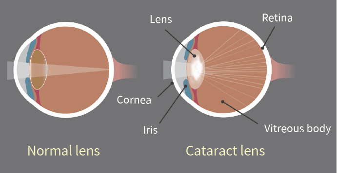
Bioguard Corporation Canine juvenile cataract (JHC) is a hereditary form of cataract characterized by cloudiness and degeneration of the lens in the eye. This condition prevents light and images from passing through the lens to the retina, impairing the dog’s vision and eventually leading to blindness. Affected dogs may exhibit symptoms such as bumping into furniture, losing direction, and moving slowly. JHC can also lead to intraocular complications, including uveitis, glaucoma, and lens luxation. Dogs are more prone to cataracts than any other species, and while cataracts can develop at any age, juvenile cataracts typically occur in dogs under 7 years old. Genetic mutaion related to JHC JHC is caused by a mutation in the heat shock transcription factor 4 (HSF4) gene, leading to an autosomal recessive inheritance pattern. Assuming that “A” represents a normal allele and “a” a mutated allele, an individual will exhibit symptoms of the disease only if both alleles are mutated (aa). Diagnosis A comprehensive ophthalmological examination is essential to assess the severity of cataracts and identify any associated complications. This examination typically includes tests for vision, pupillary light reflex, cataract severity and grading, and checks for any concurrent eye diseases or complications. Additional tests may include intraocular pressure measurement, tear production evaluation, corneal health assessment, fundus reflex and retinal examination, and ocular ultrasound. Genetic testing can further confirm the presence of genetic defects and is valuable as a pre-breeding health check to prevent the transmission of hereditary cataracts to offspring. Affected Breeds Breeds that are particularly prone to hereditary cataracts include Poodles, Cocker Spaniels, Boston Terriers, Siberian Huskies, Karelian Bears, Wire-haired Fox Terriers, Old English Sheepdogs, Golden Retrievers, and Labradors. It is recommended to conduct genetic testing before breeding these dogs to reduce the risk of producing affected offspring. Stages Cataracts can be classified into four stages based on the degree of lens opacity: Initial Stage: A distinct opaque white spot appears in the center of the pupil, but vision remains unaffected. Immature Stage: The lens begins to thicken both in the front and back, showing partial cloudiness. Vision becomes blurry, especially in low-light conditions, although some vision is still retained. Mature Stage: The entire lens becomes fully opaque and thickened, resulting in complete vision loss. Hypermature Stage: The clouded lens begins to shrink and clear up, making this stage prone to additional complications such as uveitis, glaucoma, and severe intraocular inflammation. This stage may also involve lens dislocation or fibrosis of the posterior capsule. Impacts and Complications The most noticeable early sign of cataracts is a change in eye color, where the lens begins to appear cloudy and white. It’s important to distinguish this from nuclear sclerosis, a common condition in older dogs. As the lens becomes cloudier, a dog’s vision deteriorates, leading to unintentional collisions with objects and increased sensitivity to the environment, often giving the dog a distant or blank stare. Cataracts typically worsen over time, resulting in a range of eye-related complications. These can include increased sensitivity, lens-induced uveitis (LIU), glaucoma, and the rupture of zonular fibers around the lens, which may cause lens dislocation or subluxation, as well as opacification of the posterior capsule. As cataracts progress to the hypermature stage, additional complications such as intraocular bleeding, retinal detachment, and further instances of glaucoma may arise. Early diagnosis and treatment are essential for managing these complications effectively and improving the overall prognosis. Diagnosis A comprehensive ophthalmological examination is essential to assess the severity of cataracts and identify any associated complications. This examination typically includes tests for vision, pupillary light reflex, cataract severity and grading, and checks for any concurrent eye diseases or complications. Additional tests may include intraocular pressure measurement, tear production evaluation, corneal health assessment, fundus reflex and retinal examination, and ocular ultrasound. Genetic testing can further confirm the presence of genetic defects and is valuable as a pre-breeding health check to prevent the transmission of hereditary cataracts to offspring. References Mellersh CS, McLaughlin B, Ahonen S, Pettitt L, Lohi H, Barnett KC. Mutation in HSF4 is associated with hereditary cataract in the Australian Shepherd. Vet Ophthalmol. 2009 Nov-Dec; 12(6):372-8. Mellersh CS, Pettitt L, Forman OP, Vaudin M, Barnett KC. Identification of mutations in HSF4 in dogs of three different breeds with hereditary cataracts. Vet Ophthalmol. 2006 Sep-Oct; 9(5):369-78.
Cryptosporidiosis

Bioguard Corporation Cryptosporidiosis is an illness you get from the parasite Cryptosporidium. It causes watery diarrhea and other gastrointestinal (gut) symptoms. In addition to stomach infection, this parasite can infect the respiratory system causing a cough and/or problems breathing. The family Cryptosporididae belongs to the phylum Apicomplexa characterized by an anterior (or apical) polar complex (with apical rings, micronemes, and subpellicular microtubules), which allows penetration into host cells. Cryptosporidium species are able to infect a broad range of hosts including humans, domestic and wild animals (mammals, birds, fish, marsupials, reptiles, and amphibians) worldwide. Transmission and Life Cycle Humans and animals become infected with Cryptosporidium by touching anything that has come in contact with contaminated feces, although the most common mode of transmission is represented by ingestion of oocysts in contaminated food and water or air. Cryptosporidium has three developmental stages: meronts, gamonts, and oocysts. They reproduce within the intestinal epithelial cells. Two types of oocysts, thick-walled and thin-walled, are produced during sexual reproduction. Thick-walled oocysts are excreted from the host into the environment, whereas thin-walled oocysts are involved in the internal autoinfective cycle and are not recovered from stools. Oocysts are infectious upon excretion, thus enabling direct and immediate fecal-oral transmission. Clinical Symptoms The most common symptoms of cryptosporidiosis are watery diarrhea and stomach cramps. Other symptoms may include fever, nausea, vomiting, and loss of appetite. Symptoms and severity of infection vary with the age and immune status of the host. Cryptosporidium infections are uncommonly detected in cats and dogs. Cryptosporidiosis can sometimes make dogs and cats sick, but animals with signs are atypical. In most cases, epithelial damage is minimal, but in severe cases, infection is associated with losing the ability to maintain water balance. Clinical signs are usually restricted to mild diarrhea unless the host is immunosuppressed or has another underlying condition such as viral infection or malignancy. Diagnosis Cryptosporidiosis is a diarrheal disease that is spread through contact with the stool of an infected person or animal. The disease is diagnosed by examining stool samples. Oocyst excretion is intermittent, and multiple stool samples may be needed. Diagnostic methods include: Microscopic examination: Typically, stool samples are analyzed microscopically using various techniques, including acid-fast staining and Ziehl-Nielsen staining. Real-time PCR: The most accurate method for detecting Cryptosporidium spp. is through a fecal PCR assay. Immunologic tests: These include direct fluorescent antibody tests and enzyme immunoassays to detect Cryptosporidium sp. antigens. Treatment and Prevention Most patients with healthy immune systems will recover from cryptosporidiosis without treatment. Supportive measures, oral or parenteral rehydration, and hyperalimentation may be needed for immunocompromised patients with severe disease. The best way to prevent the spread of Cryptosporidium at home is by practicing good hygiene. References Sardinha-Silva A, Alves-Ferreira EVC, Grigg ME. Intestinal immune responses to commensal and pathogenic protozoa. Front Immunol. 2022 Sep 16;13:963723. Sponseller JK, Griffiths JK, Tzipori S. The evolution of respiratory Cryptosporidiosis: evidence for transmission by inhalation. Clin Microbiol Rev. 2014 Jul;27(3):575-86. Watier-Grillot S, Costa D, Petit C, et al. Cryptosporidiosis outbreaks linked to the public water supply in a military camp, France. PLoS Negl Trop Dis. 2022 Sep 12;16(9):e0010776.
Lyme Disease in Dogs
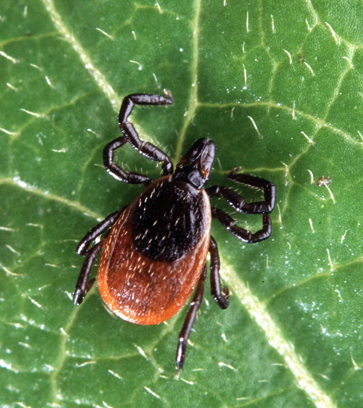
Oliver Organista, LA Lyme disease is triggered by the bacterium Borrelia burgdorferi, which belongs to the spirochete class, characterized by its worm-like, spiral shape within the genus Borrelia. This bacterium is spread to both dogs and humans through the bite of an infected black legged tick, also known as the deer tick (Ixodes scapularis). The lifecycle of the I. scapularis tick occurs at various times throughout the year, influenced by geographic location, which in turn affects the timing of Lyme disease transmission. This disease is predominantly found in certain regions, including southern New England, the eastern Mid-Atlantic, the upper Midwest (notably Wisconsin and Minnesota), and parts of the West Coast such as northern California in the United States. Lyme disease is also encountered in Europe and Asia. Typical habitats for these ticks include forests, grassy areas, wooded and marshy zones near bodies of water, and secluded or rural parts of homes and buildings. In Canada, Lyme disease can be spread by two types of black legged ticks: Ixodes scapularis and Ixodes pacificus. Dogs are most often bitten by adult I. scapularis ticks, which are particularly active during the cooler months of early spring and late fall. It is rare for a female tick to pass B. burgdorferi directly to her offspring. Ticks generally acquire the infection during their juvenile stages after feeding on an infected wildlife host, typically rodents. Since ticks feed only once at each life stage, the bacterium’s next chance to spread occurs with the tick’s subsequent blood meal in its next developmental stage. Clinical symptoms Symptoms of Lyme disease typically emerge several months after infection, often between two to five months post-exposure. The clinical presentation of Lyme disease can closely resemble that of anaplasmosis, as both diseases share similar symptoms and occur in similar regions of the country. The most common indicators of Lyme disease in dogs include: Lameness: One of the hallmark signs of Lyme disease is the inability to properly use one or more limbs, often due to pain. Swollen lymph nodes: The swelling of lymph nodes, located in areas such as the neck, chest, armpits, groin, and behind the knees, often signals an immune response to the infection. Joint swelling: Dogs may show signs of swollen joints, which can lead to stiff movements or reluctance to be touched, indicative of discomfort. Fatigue: Affected dogs may display flu-like symptoms, including a noticeable decrease in energy and increased lethargy. Loss of appetite: A reduction in eating habits, particularly if it results in weight loss, can be a symptom of Lyme disease. Fever: A fever is another possible symptom that can accompany the other signs mentioned. In some uncommon instances, untreated Lyme disease can lead to serious complications involving the kidneys, nervous system, and heart. Kidney involvement is the second most frequent severe manifestation of Lyme disease in dogs and often proves to be fatal. Cases involving the nervous system may present with facial paralysis and seizures. Heart-related complications, while rarer, have also been documented. Diagnosis To diagnose Lyme disease in dogs, serologic assays are the most widely used methods. While some laboratories continue to utilize traditional approaches like the whole-cell enzyme-linked immunosorbent assay (ELISA) , the immunofluorescence assay (IFA), and rapid tests. These tests identify the presence of antibodies against specific proteins of B. burgdorferi, offering a simple yes/no result regarding the dog’s serology status. Treatment Treatment is advised for dogs that test positive for Lyme disease and show clinical symptoms, as well as for asymptomatic dogs that have signs of protein-losing nephropathy. The antibiotics doxycycline and minocycline are the primary medications used, administered at a dose of 10mg/kg orally every 12 to 24 hours for 30 days. Amoxicillin and erythromycin are alternative antibiotic options. Additionally, a non-steroidal anti-inflammatory drug (NSAID), such as carprofen or deracoxib, may be prescribed to manage symptoms. Prevention To safeguard against Lyme disease, the most effective approach is the consistent use of tick-prevention products throughout the year. There are numerous commercial options available for controlling ticks on both dogs and cats, such as systemic treatments (like isoxazolines), topical applications (such as permethrin and fipronil), and tick-preventive collars. Vaccination also serves as an effective means of protection. Additionally, limiting exposure to tick-infested areas and practicing caution in environments known to harbor ticks are important preventive strategies. References 2. Lyme Disease in Dogs: Signs and Prevention, Kathryn E. Reif, MSPH, PhD.,April 2020, https://todaysveterinarypractice.com/parasitology/lyme-disease/ 3. Lyme Disease, IPAC (https://ipac-canada.org/lyme-disease.php) 4. Littman MP, Gerber B, Goldstein RE, et al. ACVIM consensus update on Lyme borreliosis in dogs and cats. J Vet Intern Med 2018;32(3):887-903. 5. Mullegger RR. Dermatological manifestations of Lyme borreliosis. Eur J Dermatol. 2004 Sep-Oct;14(5):296-309. PMID: 15358567