
Long Pham
Introduction
Psittacine beak and feather disease (PBFD) is an infectious viral disease that infects psittacine birds. This disease affects Old World (Australian and African) psittacine birds and New World (Americas) psittacine birds (Greenacre, 2005). The peracute and acute form of this disease can cause sudden death, while the chronic form of this disease damages the feather, deforms the beak, and will eventually lead to death. (KATOH et al., 2010)
The disease is caused by a small circovirus, which is a single-stranded DNA virus belonging to the Circoviridae family (Hakimuddin et al., 2016). The virus can spreads through direct contact with contaminated surfaces, feces, feather dander, and other bodily excretions (Greenacre, 2005). It can be transmitted horizontally to other birds in the same generation and vertically to eggs and young chicks in the next generation (Hakimuddin et al., 2016). Since the virus has a non-envelope structure, it is able to resist many control measures and is able to persist in the environment and infected substances for a long time.
The origin of PBFD was thought of to be from Australia (PASS & PERRY, 1984), where it then spread to the rest of the world. Possibly through pet trades and import of these birds, this disease was able to spread globally. Report of this disease has occurred in other countries located in North America, Europe, Africa, Asia, and even on islands in the Indian and Pacific Oceans (Harkins et al., 2014). Psittacine beak and feather disease prevalence around the world varies and has been reported to be around 41.2% in Taiwan (Hsu et al., 2006), 3.5–4% in USA (de Kloet & de Kloet, 2004) and 23% in Australia (Khalesi et al., 2005). With an increasing trend of live birds being traded globally, the spread of PBFD and other diseases will surely grow.
Diagnosis
Typical clinical signs of PBFD include lethargy, weight loss, shedding and abnormal development of feathers, beak elongation and deformation, and eventually death (PASS & PERRY, 1984). This disease can occur in three different forms: peracute, acute, and chronic. Progression of the disease depends on the age, with younger birds having a higher progression rate (Greenacre, 2005).
Some symptoms of peracute PBFD are weight loss, pneumonia, sepsis, enteritis, liver necrosis, and leukopenia (Schoemaker et al., 2000). Sudden death is likely to occur in peracute PBFD.
In acute PBFD, majority of those affected by this phase are between the ages of 0-3 years old and it is thought that their susceptibility is based on their condition instead of the virus’ antigenic or genotypic characteristics (Ritchie et al., 1990). Some clinical signs includes depression and rapidly developing feather dystrophy can occur, affecting 80-100% of the feathers in as little as one week (Ritchie, 1995). Sudden death can also occur in this form. Those that survive this phase will have an incubation period, which may be years, before going to the chronic PBFD phase (Greenacre, 2005).
For chronic PBFD, it is typically characterized by symmetrical feather dystrophy that progresses slowly and gets worse over time (Greenacre, 2005). Birds can become completely bald and can have beak deformities (Figure 1), where the beak becomes elongated. Death usually occurs from secondary infections, fungal or bacterial, because lymphoid tissues are usually damaged by the virus and causes the immune system to be suppressed (Ritchie et al., 2003).
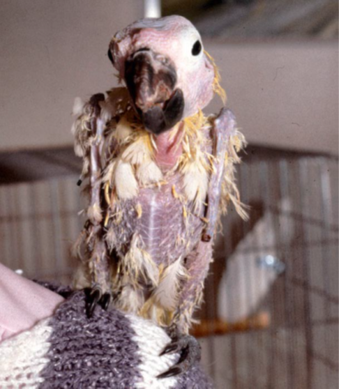
Figure 1: Cockatoo with advanced PBFD (Harcourt-Brown, 2009)
PBFD can be diagnosed successfully from just careful examination. The disease can first be suspected if the bird is progressively losing feathers or has a symmetrical feather dysplasia. However, a loss of feathers does not always mean it is PBFD as the cause can be from other reasons, such as being self-inflicted or from excessive allopreening, which causes injuries that look similar to those caused by the disease (Wellehan et al., 2016). PBFD can be diagnosed though antigen and antibody detection from hemagglutination assay and hemagglutination inhibition. In addition, polymerase chain reaction (PCR) is also used for detecting PBFD, being a standard method of detection in most countries (Wellehan et al., 2016). False positives can occur with this method due to the nature of the virus to easily contaminate and persist in the environment, which will contaminate the samples, such as feathers, that are exposed to this environment (Wellehan et al., 2016). Therefore, the choice of sample collection method can have a major impact to the results. In one study, it was found that the use of blood samples for a PCR test resulted in 47 out 56 birds being positive for PBFD, while only 10 birds had a positive result when feather samples were used (Khalesi et al., 2005).
Treatment and Disease Control
Current treatment for PBFD is for supportive care to prevent secondary infections as there is no cure for this disease. The disease is fatal when clinical signs appear, while other birds that has an immune response and don’t show any clinical signs, making this naturally vaccinated (Greenacre, 2005). Effective methods of controlling this disease involves isolating suspected carriers, testing, and if necessary, culling to prevent a possible outbreak from occurring. The resilience of the virus to many chemical disinfectants and even extreme temperatures can be based on the physicochemical properties of the virus (Raidal & M.Cross, 1994). However, Virkson S or other peroxide disinfectants have been suggested for use to disinfect contaminated areas (Wellehan et al., 2016). Strict hygiene practices with the right disinfectants is the key to prevent further spread of PBFD. While there are no vaccine for PBFD available commercially, research into developing one is ongoing and currently made vaccines appears to be effective. Future control of the disease will still depend on implementing strict hygiene practices and testing methods since vaccinated birds may still spread the disease.
References
Greenacre, C.B. (2005) Viral diseases of companion birds. Veterinary Clinics of North America: Exotic Animal Practice, 8, 85–105.
KATOH, H., OGAWA, H., OHYA, K. & FUKUSHI, H. (2010) A review of DNA viral infections in Psittacine birds. Journal of Veterinary Medical Science, 72, 1099–1106.
Hakimuddin, F., Abidi, F., Jafer, O., Li, C., Wernery, U., Hebel, C. & Khazanehdari, K. (2016) Incidence and detection of beak and feather disease virus in psittacine birds in the UAE. Biomolecular Detection and Quantification, 6, 27–32.
PASS, D.A. & PERRY, R.A. (1984) The pathology of Psittacine Beak and feather disease. Australian Veterinary Journal, 61, 69–74.
Harkins, G.W., Martin, D.P., Christoffels, A. & Varsani, A. (2014) Towards inferring the global movement of beak and feather disease virus. Virology, 450-451, 24–33.
Hsu, C.-M., Ko, C.-Y. & Tsai, H.-J. (2006) Detection and sequence analysis of avian polyomavirus and psittacine beak and feather disease virus from Psittacine birds in Taiwan. Avian Diseases, 50, 348–353.
de Kloet, E. & de Kloet, S. (2004) Analysis of the beak and feather disease viral genome indicates the existence of several genotypes which have a complex psittacine host specificity. Archives of Virology, 149, 2393–2412.
Khalesi, B., Bonne, N., Stewart, M., Sharp, M. & Raidal, S. (2005) A comparison of haemagglutination, haemagglutination inhibition and PCR for the detection of psittacine beak and feather disease virus infection and a comparison of isolates obtained from Loriids. Journal of General Virology, 86, 3039–3046.
Schoemaker, N.J., Dorrestein, G.M., Latimer, K.S., Lumeij, J.T., Kik, M.J., van der Hage, M.H. & Campagnoli, R.P. (2000) Severe leukopenia and liver necrosis in young African grey parrots (Psittacus erithacus erithacus) infected with Psittacine Circovirus. Avian Diseases, 44, 470.
Ritchie, B.W., Niagro, F.D., Latimer, K.S., Lukert, P.D., Steffens, W.L., Rakich, P.M. & Pritchard, N. (1990) Ultrastructural, protein composition, and antigenic comparison of psittacine beak and feather disease virus purified from four genera of Psittacine Birds. Journal of Wildlife Diseases, 26, 196–203.
Ritchie, B.W. (1995) Avian viruses, function and control. Wingers Publishing.
Ritchie, P.A., Anderson, I.L. & Lambert, D.M. (2003) Evidence for specificity of psittacine beak and feather disease viruses among avian hosts. Virology, 306, 109–115.
Harcourt-Brown, N. (2009) Psittacine Birds. Handbook of Avian Medicine, pp. 138-168.
Wellehan, J.F.X., Lierz, M., Phalen, D., Raidal, S., Styles, D.K., Crosta, L., Melillo, A., Schnitzer, P., Lennox, A. & Lumeij, J.T. (2016) CHAPTER 2 – Infectious disease. Current therapy in Avian Medicine and Surgery (ed B.L. Speer), pp. 22–106.