
Bioguard Corporation
Pigeon circovirus (PiCV) is an infectious disease that mainly affects young pigeons between 1 and 4 months of age. Mortality is variable, but it can approach 100%. Pigeon circovirus infections have led to the loss of lymphoid tissue in immune system organs, and for this reason, PiCV is regarded as an immunosuppressive agent in pigeons.
Etiology
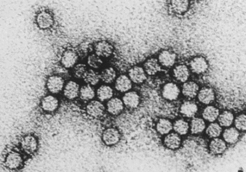
Pigeon circoviruses, also known as columbid circovirus (CoCV), belong to the genus Circovirus in the family of the Circoviridae. They are small, non-enveloped viruses consisting of single-stranded circular DNA. Pigeon circovirus is antigenically distinct from the psittacine beak and feather disease (PBFD) virus, which also belongs to the genus Circovirus, but does share some homologous.
Transmission
The pigeon circovirus is transmitted mainly horizontally. Detection of the virus in the intestine, cloaca, and feces support fecal–oral route transmission. Also, inhalation of other fecal-contaminated materials, such as feather dust, has been suggested as a potential respiratory route of infection. Vertical transmission of PiCV is also possible by the detection of the virus in testis and semen samples of breeding cocks, the ovary but not in the oviduct of the hens, in embryonated eggs, and chicks recovered from eggs shortly before hatching.
Clinical signs and lesions
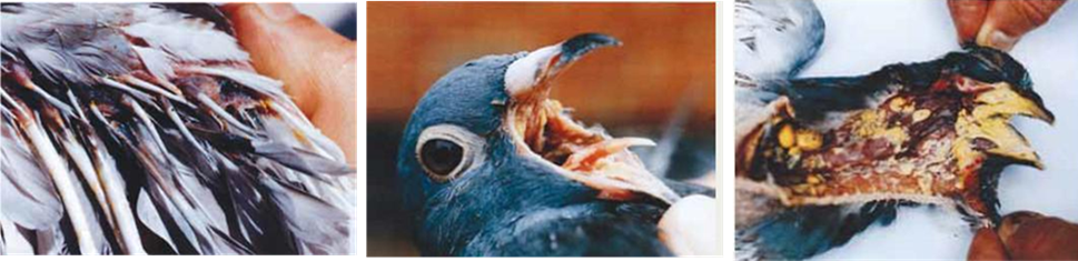
The clinical signs of PiCV infections in pigeons is highly varied. In pigeons between 1 and 4 months of age, circovirus infections are associated with lethargy, anorexia, ruffled feathers, dyspnea, diarrhea, and fluid-filled crop, also known as ‘young pigeon disease syndrome’ (YPDS). However, this syndrome should be considered a multifactorial disease. In most cases, concurrent infections can be demonstrated with all possible viral, bacterial, and parasitic agents hampering the assessment of the exact role of the circovirus.
The most obvious lesion is a swollen, edematous bursa in the acute phase of infection. However, more chronic infections result in atrophy of the bursa. Histological lesions consist of lymphocyte depletion in lymphoid tissue and characteristic intracytoplasmic basophilic inclusion bodies (mainly in macrophages) in the lymphoid tissue.
Diagnosis and Prevention
Before having the sequence of the pigeon circovirus, diagnosis of PiCV infections mainly depended on histopathological examinations and electron microscopy findings of the intracytoplasmic and/or intranuclear inclusion bodies in lymphoreticular and hepato-intestinal tissues. Since publication of PiCV sequence, the virus now can be detected by polymerase chain reaction (PCR), in situ hybridization, and dot blot analysis. Currently, there is no specific treatment or effective vaccine against PiCV since pigeons infected with PiCV could die from secondary infections caused by the virus. At the moment, protection against potential detrimental effects of PiCV infection relies on good biosecurity measures in the loft.
Reference
1. Schmidt R.E. Circovirus in Pigeons. J. Assoc. Avian Vet. 1992;6:204.
2. Woods L.W., Latimer K.S., Niagro F.D., Riddell C., Crowley A.M., Anderson M.L., Daft B.M., Moore J.D., Campagnoli R.P., Nordhausen R.W. A retrospective study of circovirus infection in pigeons: Nine cases (1986–1993) J. Vet. Diagn. Investig. 1994;6:156–164.
3. Tian D. Bibliometric analysis of pathogenic organisms. Biosaf. Health. 2020;2:95–103.
