
An Overview Of Feline Parvovirus
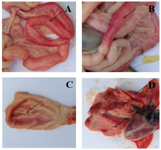
Trinh Mai Nguyen Tang Feline panleukopenia virus (FPV) is a single-stranded DNA virus that causes feline leukopenia disease. It belongs to the family Parvoviridae, and is also known as Parvo in cats [1]. In the early years of the twentieth century, the first cases of FPV were reported in cats [2]. This is a common and dangerous disease in cats worldwide that occurs seasonally, especially from early summer to autumn with the highest peak in July, August, and September [3]. The virus usually resides in the everyday items of cats such as cages, beds, food bowls, etc [4]. Moreover, it has high resistance to physical factors and disinfectants, they can still survive for 30 minutes at temperatures up to 560C, and can even survive in contaminated environments for months or even years, which is why FPV is highly lethal and contagious in cats [4-5]. Transmission and clinical signs FPV is spread by the fecal-oral route when cats come into contact with food, water, objects, or secretions that contain the virus. The virus enters and infects cells through the feline transferrin receptor (fTfR) [6-7]. Previous reports also showed that the two main types of cells that FPV replicate are small intestinal crypt cells and lymphoid cells [8-9]. The replication of virus leads to impaired white blood cell function, and bleeding in the small intestine and stomach is the cause of hemorrhagic diarrhea in cats [10]. According to Yen et al.’s report in 2021, the percentage of small intestine congested is 100% in a total of 8 cats examined (Figure 1A-B), and 75% of intestinal mucosal ulcerations appear. Meanwhile, there are 6 cats with gastric congestion in 75% (Figure 1C) and 2 cats with pneumonia accounting for 25% (Figure 1-D). In addition, other lesions were also detected, such as enlarged lymph nodes in the mesentery, and infarcted spleen [10]. These lesions can cause symptoms such as anorexia, fever, vomiting, and hair loss in the early stages [11], followed by diarrhea, severe dehydration, electrolyte imbalance, hypoglycemia, hemorrhage or sepsis and endotoxins in the blood cause rapid death in cats if not treated promptly [12-15]. Furthermore, in some kittens, there is damage to the central nervous system such as hydrocephalus, and cerebellar hypoplasia [16]. According to Reif (1976), cats infected with FPV have an incubation period of 4 to 6 days [16], during infection can excrete virus-containing feces for up to 43 days, and they even can survive for more than 1 year in the lungs and kidneys [17] Figure 1. Symptoms of cat infection with FPV. A, B: Intestinal congestion, scattered or intermittent bleeding; C: Congested stomach contains a lot of fluid; D: Mild pneumonia [10] FPV can cause infection in cats 3 to 5 months of age when infected with them or in unvaccinated cats, especially kittens, with an almost 90% chance of mortality due to maternally derived antibodies (MDAs) weakened [18]. MDAs may protect kittens against disease during the first weeks of life, and they are mainly transmitted through colostrum [19-20]. However, a kitten’s immunity will depend on factors such as the length of the lactation, the quality of the colostrum, and the amount of milk they are ingested [21]. Figure 2. The immunity gap and maternally derived antibodies (MDAs) [24] Although kittens all have antibodies inherited from the mother, these antibodies can only protect for 6-8 weeks, these antibodies gradually disappear and create an immunity gap that makes it easier for the virus to attack [19]. The immunological gap (Figure 2) is the distance that the maternally derived antibodies from the mother cat to the kittens are reduced. This gap usually occurs when kittens reach the age of 8-12 weeks and interferes with the development of immunization immunity [24]. There is evidence that there is seroconversion with long-term antibody titers after administration of modified live vaccines (MLV) in kittens lacking MDAs compared with kittens receiving MDAs [22–23]. When MDA titres ≥1:10 as measured by inhibition of coagulation (HI), MLV vaccination should not be given because of seroconversion [19, 24]. Diagnosis FPV can be difficult to diagnose accurately because it has similar symptoms to feline immunodeficiency virus (FIV) infection. Symptoms of feline leukemia virus (FeLV) infection and pancreatitis vary depending on the extent of the infection. FPV detection using PCR There are many methods to detect the presence of FPV in the cat. One of the most widely used and informative methods today is PCR testing, which can use whole blood samples or feces [26]. FPV detection using ELISA method or Rapid test kit The ELISA method can detect the presence of FPV-specific antigens in cat feces. When cats show clinical symptoms, they can be tested by ELISA or using the FPV antigen rapid test kit. The FPV Antigen test kit made by Bioguard Corporation is simple, fast, and accurate with a specificity and sensitivity of 92.54%. FPV detection using blood test method One of the methods of FPV diagnosing is to count white blood cells through a blood test. Other reports have also shown that markedly reduced leukocyte counts in hematologic parameters are very common in cats with FPV usually below 3000 and may reach less than 200/mm3 [3]. Prevention and Treatment Because the FPV virus can survive for a long time in the environment and has high resistance to physical factors and disinfectants so it is necessary to clean the barn, shelter, and food bowl after the cat has been infected. In addition, it is necessary to isolate infected cats to avoid infecting other cats and becoming an outbreak FPV in cats is a dangerous disease with no cure. All treatment for leukopenia in cats is for symptomatic treatment. Although there is no cure for feline leukopenia, if detected in time, the symptoms can be treated and the cat can recover. Prevention The way to prevent cats from contracting FPV is vaccination. Atteunuated live virus vaccines can be used, but should not be given to kittens < 4 weeks of