Canine Distemper Virus
Tang Nguyen Mai Trinh
Infection of canine distemper virus (CDV) is common in most terrestrial carnivores in the wild, especially in the Canidae family (eg, wolves, foxes, domestic dogs, etc.), as well as certain ferrets, otters, raccoons, cats, and even marine animals [1]. CDV is a member of the Paramyxoviridae family which the genus Morbilivirus was first described in the 1970s, however, it was only isolated in 1905 from the nasal secretions of infected dogs by a French veterinarian, [2]. CDV is now known as the most dangerous disease in dogs because of its propensity to infect through respiratory and digestive pathways and cause death in virtually all canines. When infected with it, the unvaccinated host has a 100% mortality rate [3].
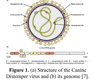
Structure of Canine distemper virus
CDV is known as a single-stranded RNA virus, like many other viruses CDV has an N nucleocapsid protein that is responsible for genome replication, matrix (M), fusion (F), phosphoprotein (P), and polymerase (L) which are involved in genomic and mRNA transcription. Whereas nonstructural proteins C and V have been identified as virulence factors involved in the modulation of immune responses and innate immunity, proteins like H and F are essential for viral attachment, invasion, and fusion into host cells [4-6].
Transmission
Puppies and dogs most often become infected through airborne exposure to the virus from an infected dog or wild animal. CDV enter the dog’s body through respiratory secretions, most notably coughing and sneezing. Virus particles can spread about 25 feet with a single sneeze, then proliferate in immune cells such as macrophages, T and B cells via the SLAM receptor within 24 hours [8] and spreads to tonsils and lymph nodes via the lymphatic vessels, causing long-term immunosuppression [9-11]. The amount of virus in the lymph nodes of the throat, tonsils, and bronchi will go up after 2 to 4 days of infection, and the virus continues to release and replicate in multiple organs. This process takes 4 to 6 days after infection and affects organs such as the thymus, spleen, lymph nodes, stomach cells, lungs, and bone marrow. The increased concentration of virus in the lymph node tissue at this time causes the number of white blood cells to decrease, resulting in the infected dog’s body temperature increasing [12]. The virus begins to migrate to specific epithelium and the central nervous system (CNS) via the blood or CSF after around 8 –10 days [10]. Simultaneously, organs such as the respiratory tract, intestines, and urinary tract begin to experience significant infections as lymphocytes are destroyed, resulting in a massive release of viruses into these organs [13].
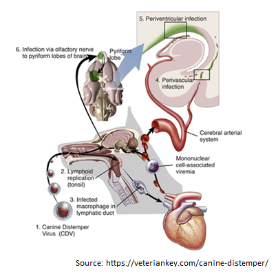
Figure 2. The process of entry and replication of CDV in dogs.1, The CDV virus enters the dog’s body through the respiratory tract. 2, the virus begins to multiply in the tonsils, bronchial lymph nodes, oropharyngeal nodes, and gastrointestinal lymphoid tissues. 3, the virus attacks immune cells such as macrophages and is released to other organs such as the heart. 4, The virus enters the central nervous system (CNS) through the circulatory system. 5, Virus enters the cerebrospinal fluid (CFS). 6, Viral migration to the pyramidal lobes of the cerebral cortex through the nasal passages to the central nervous system [14].
Clinical Symptoms of Canine Distemper Virus infection in dogs
The CDV virus can cause disease in dogs of any age, although it is more common in pups aged 12 to 16 weeks and in unvaccinated dogs. Because pups’ immune systems are so immature, they are easily infected by the virus if they come into touch with dogs that have already been afflicted with CDV. When the dog get infected with the CDV virus, early symptoms include fever, weariness, lack of appetite, apathy, and a reluctance to be active [9]. However, these symptoms are quite similar to those of the parvovirus, which primarily infects the digestive system, whereas CDV not only assaults the digestive system but also damage to the respiratory system, neurological system, bones, arthritis, eyes, and skin [13].
Prolonged vomiting, diarrhea, severe dehydration, and stool abnormalities in color, consistency, and blood and mucus contribute to gastritis and intestinal inflammation in the early stages. Finally, the dog was unable to defecate, resulting in death (Figure 3A). Pus discharge, ulcers, and progressively becoming clouded eyes can lead to conjunctivitis and even blindness, while the skin appears as spots with pus within, then gradually changes from red to yellow (Figures 3 B and C). Signs such as a runny nose with green mucus in the early stage, shortness of breath in the latter stage, and shortness of breath lead to pneumonia in the respiratory system (Figure 3B).
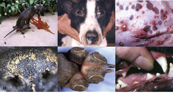
Figure 3. Signs of CDV infection in dogs. A, Dogs have digestive system damage, diarrhea with blood [17]. B, Eyes are damaged causing pus discharge, ulcers, while the nose shows signs of runny nose with green mucus [15]. C, The skin also appears spots that have pus inside. D and E, the dog’s paw and nose epithelium show basal keratinocytes. F, enamel deterioration in adult dogs following neonatal CDV infection [13].
When the disease is severe, evidence of central nervous system damage such as convulsions, hemiplegia, or quadriplegia will occur; at this stage, 99% of infected dogs will die. Furthermore, the CDV virus can attach to lymphocytes and enter the cerebrospinal fluid (CSF), causing harm to the central nervous system. In addition, CDV was discovered in the foot and nasal epithelium, promoting basal keratinocyte growth, which was clearly recognized in the Figures 3D and E.
Although CDV infection leads to the above clinical signs, however the appearance of these signs is also dependent on habitat, age, host immunity, and virulence of the virus strain [13]. Studies have also demonstrated that CDV can be passed from mother to puppy through the placenta during pregnancy. When the mother dog is infected with the virus, it can lead to miscarriage, stillbirth, but this depends on the pregnancy stage of the mother dog [16]. For the lucky puppies born, due to damage to the lymphatic system during infection, they will be permanently immunocompromised. In puppies, before the eruption of permanent teeth, infection with CDV can cause damage to the roots and enamel (Figure 3F).
How to detect Canine distemper virus disease in dog?
Most dogs infected with CDV are discovered in the late stages of the disease, and they are unable to be treated. For testing CDV infections, some methods can be used such as real-time PCR, ELISA, etc,
As for the Real time PCR method, it amplify the presence of RNA with high accuracy, allowing the detection of small amounts of virus in the sample. Clinical samples such as whole blood, conjunctiva, urine or feces, which contain high levels of virus. However, in dogs that have been vaccinated for about 2 weeks, PCR testing should not be performed because of the high probability of false-positives caused by the modified virus from the vaccine [18].
Enzyme-Linked Immunosorbent Assay, which detects the presence of CDV in serum and plasma samples by measuring levels of IgM (generated in the early stages of infection) and “IgG” (made in the early stages of infection) [19]. This method is simple, fast, specific and sensitive. This consistently high IgM level indicates infection caused by CDV or recent vaccination, however this cannot demonstrate clinical disease [19-20].
In addition, other methods such as rapid test kit, immunofluorescence (IF) [21, serology test (SN) [22] can also be used in the diagnosis of infection caused by CDV.
References
- Mayer, J., & Donnelly, T. M. (2012).Clinical veterinary advisor: birds and exotic pets. Elsevier Health Sciences).
- Carré H. Sur la maladie des jeunes chiens. C R Acad Sci. 1905;140(689–690):1489–1491.
- Patel, J. R., Heldens, J. G. M., Bakonyi, T., & Rusvai, M. (2012). Important mammalian veterinary viral immunodiseases and their control.Vaccine, 30(10), 1767-1781.
- Cattaneo, R., Kaelin, K., Baczko, K., & Billeter, M. A. (1989). Measles virus editing provides an additional cysteine-rich protein.Cell, 56(5), 759-764.
- Yang, D. K., Kim, H. H., Lee, S., Yoon, Y. S., Park, J., Oh, D., … & Hyun, B. H. (2020). Isolation and molecular characterizations of canine distemper virus from a naturally infected Korean dog using Vero cells expressing dog signaling lymphocyte activation molecule.Journal of Veterinary Science, 21(5).
- Nakatsu, Y., Takeda, M., Ohno, S., Shirogane, Y., Iwasaki, M., & Yanagi, Y. (2008). Measles virus circumvents the host interferon response by different actions of the C and V proteins.Journal of virology, 82(17), 8296-8306.
- Rosado, R. C. (2009).Rastreio virológico de carnívoros errantes e caracterização genética viral (Bachelor’s thesis, Universidade Técnica de Lisboa. Faculdade de Medicina Veterinária).
- Seki, F., Ono, N., Yamaguchi, R., & Yanagi, Y. (2003). Efficient isolation of wild strains of canine distemper virus in Vero cells expressing canine SLAM (CD150) and their adaptability to marmoset B95a cells. Journal of virology, 77(18), 9943-9950.
- Vandevelde, M., & Zurbriggen, A. (1995). The neurobiology of canine distemper virus infection. Veterinary Microbiology, 44(2-4), 271-280.
- Krakowka, S. (1982). Mechanisms of in vitro immunosuppression in canine distemper virus infection. Journal of Clinical & Laboratory Immunology, 8(3), 187-196.
- Krakowka, S., Higgins, R. J., & Koestner, A. (1980). Canine distemper virus: review of structural and functional modulations in lymphoid tissues. American Journal of Veterinary Research, 41(2), 284-292.
- Tipold, A., Vandevelde, M., & Jaggy, A. (1992). Neurological manifestations of canine distemper virus infection. Journal of Small Animal Practice, 33(10), 466-470.
- von Messling, D. Milosevic, & R. Cattaneo. (2004) “Tropism illuminated: lymphocyte-based pathways blazed by lethal morbillivirus through the host immune system,” Proceedings of the National Academy of Sciences of the United States of America, vol. 101, no. 39, pp. 14216–14221.
- Craig E. Greene & Marc Vandevelde. Canine Distemper, chapter 3, https://veteriankey.com/canine-distemper/
- https://www.marvistavet.com/canine-distemper.pml
- Krakowka, S., Hoover, E. A., Koestner, A., & Ketring, K. (1977). Experimental and naturally occurring transplacental transmission of canine distemper virus. American journal of veterinary research, 38(7), 919-922.
- http://marphavet.com/vi/news/Benh-Dieu-Tri/Benh-Care-o-cho-Benh-sai-sot-350/
- Elia, G., Decaro, N., Martella, V., Cirone, F., Lucente, M. S., Lorusso, E., … & Buonavoglia, C. (2006). Detection of canine distemper virus in dogs by real-time RT-PCR. Journal of virological methods, 136(1-2), 171-176.
- Day, M. J., & Washabau, R. J. (Eds.). (2013). Canine & Feline Gastroenterology. pp 973-996.
- Von Messling, V., Harder, T. C., Moennig, V., Rautenberg, P., Nolte, I., & Haas, L. (1999). Rapid and sensitive detection of immunoglobulin M (IgM) and IgG antibodies against canine distemper virus by a new recombinant nucleocapsid protein-based enzyme-linked immunosorbent assay. Journal of Clinical Microbiology, 37(4), 1049-1056.
- Jóźwik, A., & Frymus, T. (2005). Comparison of the immunofluorescence assay with RT-PCR and nested PCR in the diagnosis of canine distemper. Veterinary research communications, 29(4), 347-359.
Costa, V. G. D., Saivish, M. V., Rodrigues, R. L., Lima Silva, R. F. D., Moreli, M. L., & Krüger, R. H. (2019). Molecular and serological surveys of canine distemper virus: A meta-analysis of cross-sectional studies. PloS one, 14(5), e0217594.