
Maigan Espinili Maruquin
- The Feline Infectious Peritonitis (FIP)
The coronaviruses are enveloped, positive-sense single-stranded RNA viruses with non-segmented genomes of around 30,000 nucleotides in length (Tasker 2018)( Siddell SG, 1995). The feline coronavirus (FCoV) has two pathotypes distinguished by their biological behavior. The highly prevalent feline enteric coronavirus (FECV) is highly contagious with transmission from faeces of shedding cats (Felten and Hartmann 2019). However, most cases are asymptomatic or displays mild gastrointestinal clinical signs, (Addie, Toth et al. 1995, Pedersen, Sato et al. 2004, Pedersen, Allen et al. 2008, Pedersen 2009, Vogel, Van der Lubben et al. 2010, Tasker 2018, Felten and Hartmann 2019). On the other hand, the feline infectious peritonitis virus (FIPV) is a mutation within a small percentage of infected cats and it results to a fatal disease feline infectious peritonitis (FIP), commonly in young cats (Pedersen, Boyle et al. 1981, Pedersen, Boyle et al. 1981, Addie, Toth et al. 1995, Vennema, Poland et al. 1998, Pedersen 2009, Tasker 2018, Felten and Hartmann 2019). However, the exact gene causing mutation is still unknown (Felten and Hartmann 2019).
The FIP may appear in two clinically distinct forms: the wet form and dry form, which is effusive and granulomatous forms, respectively (Wolfe and Griesemer 1966, Montali and Strandberg 1972, Pedersen 2009, Hazuchova, Held et al. 2016). The development of FIP is affected by three factors. First is the viral factor wherein studies relative to mutation of the FCoV S gene where presented (Tasker 2018) and the replication in monocytes, and activation of infected monocytes were also considered important in the development of FIP (Kipar and Meli 2014, Tasker 2018). Second factor considered is the host’s immune response, breed and genetic (de Groot-Mijnes, van Dun et al. 2005, Dewerchin, Cornelissen et al. 2005, Golovko, Lyons et al. 2013, Pedersen, Liu et al. 2016, Tasker 2018). Finally, another factor affecting the FIP development is the environment- level of stress and overcrowding (Tasker 2018).
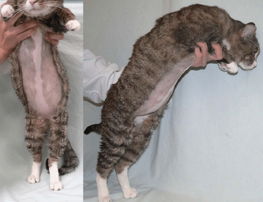
| https://www.researchgate.net/figure/Cat-with-wet-effusive-form-of-FIP-presenting-moderate-abdominal-distention-due-to_fig7_51758582 |
Fig. 01. Manifestation of moderate abdominal distention due to peritoneal effusion. A clinical sign of wet (effusive) FIP.
Common clinical signs for the FIP infected cats include lethargy, anorexia, weight loss, fluctuating pyrexia, and sometimes presents jaundice (Tasker 2018). On the other hand, wet FIP cases can be associated with abdominal, pleural and/ or pericardial effusions, and progresses within few days to weeks with severe limiting survival (Ritz, Egberink et al. 2007, Tasker 2018). Whereas, dry FIP usually displays neurological signs (Crawford, Stoll et al. 2017) or ocular signs, which progresses in a few weeks to months and are more chronic (Tasker 2018).
- The alpha- 1 acid glycoprotein (AGP) in FIP diagnosis
Prior to acquired immune response, part of the innate response is the acute phase response (Murata, Shimada et al. 2004, Schmidt and Eckersall 2015). Proteins known as the acute phase proteins (APPs) are then increased in production from hepatocytes and peripheral tissues and then released (Schmidt and Eckersall 2015). These blood proteins can be used to evaluate the innate response to infection, inflammation or trauma (Murata, Shimada et al. 2004, Petersen, Nielsen et al. 2004, Ceron, Eckersall et al. 2005, Eckersall and Bell 2010). With changes by >25% in the serum concentration in response to disease stimulation, APPs are considered useful quantitative biomarkers of diseases- in diagnosis, prognosis, response to therapy, and in general health screening (Eckersall and Bell 2010).
In response to inflammations, the serum alpha- 1 acid glycoprotein (AGP) concentration increases as a major acute phase protein in cats (Ceron, Eckersall et al. 2005, Paltrinieri 2008, Giori, Giordano et al. 2011). Studies showed increased serum AGP concentration in cats infected with FIP (Duthie, Eckersall et al. 1997, Giordano, Spagnolo et al. 2004, Giori, Giordano et al. 2011).
The feline AGP in both serum and peritoneal fluid are known biomarker for FIP (Duthie, Eckersall et al. 1997, Giordano, Spagnolo et al. 2004, Eckersall and Bell 2010). In a study conducted, AGP in effusion showed to be the best APP to distinguish between cats with and without FIP (Hazuchova, Held et al. 2016). Although AGP elevations are not specific for FIP, the measurement is helpful in the diagnosis of FIP, and levels >1.5 mg/ml are often observed in FIP cases (Tasker 2018). It was then concluded that the higher levels increase the index of suspicion (Duthie, Eckersall et al. 1997, Paltrinieri, Giordano et al. 2007, Giori, Giordano et al. 2011, Hazuchova, Held et al. 2016, Tasker 2018).
With difficulty in diagnosing FIP through conventional approaches (Addie, Paltrinieri et al. 2004, Paltrinieri 2008), samples from FIP infected cats showed AGP seemed to be associated with viral antigen and are seen present in large amounts (Paltrinieri, Giordano et al. 2004, Paltrinieri 2008). Nevertheless, AGP plays role in drug-binding, as an immunomodulatory agent, and acts as a plasma transport protein (Ceron, Eckersall et al. 2005, Ceciliani, Ceron et al. 2012, Schmidt and Eckersall 2015).
References:
Siddell SG. The coronaviridae. London: Plenum Press, 1995
Drechsler, Y., Alcaraz, A., Bossong, F., Collisson, E.W., Diniz, P. (2011), “Feline Coronavirus in Multicat Environments”. Veterinary Clinics of North America Small Animal Practice 41(6):1133-69.
Addie, D. D., S. Paltrinieri, N. C. Pedersen and s. Secong international feline coronavirus/feline infectious peritonitis (2004). “Recommendations from workshops of the second international feline coronavirus/feline infectious peritonitis symposium.” Journal of feline medicine and surgery 6(2): 125-130.
Addie, D. D., S. Toth, G. D. Murray and O. Jarrett (1995). “Risk of feline infectious peritonitis in cats naturally infected with feline coronavirus.” Am J Vet Res 56(4): 429-434.
Ceciliani, F., J. J. Ceron, P. D. Eckersall and H. Sauerwein (2012). “Acute phase proteins in ruminants.” J Proteomics 75(14): 4207-4231.
Ceron, J. J., P. D. Eckersall and S. Martýnez-Subiela (2005). “Acute phase proteins in dogs and cats: current knowledge and future perspectives.” Vet Clin Pathol 34(2): 85-99.
Crawford, A. H., A. L. Stoll, D. Sanchez-Masian, A. Shea, J. Michaels, A. R. Fraser and E. Beltran (2017). “Clinicopathologic Features and Magnetic Resonance Imaging Findings in 24 Cats With Histopathologically Confirmed Neurologic Feline Infectious Peritonitis.” J Vet Intern Med 31(5): 1477-1486.
de Groot-Mijnes, J. D. F., J. M. van Dun, R. G. van der Most and R. J. de Groot (2005). “Natural history of a recurrent feline coronavirus infection and the role of cellular immunity in survival and disease.” Journal of virology 79(2): 1036-1044.
Dewerchin, H. L., E. Cornelissen and H. J. Nauwynck (2005). “Replication of feline coronaviruses in peripheral blood monocytes.” Arch Virol 150(12): 2483-2500.
Duthie, S., P. D. Eckersall, D. D. Addie, C. E. Lawrence and O. Jarrett (1997). “Value of alpha 1-acid glycoprotein in the diagnosis of feline infectious peritonitis.” Vet Rec 141(12): 299-303.
Eckersall, P. D. and R. Bell (2010). “Acute phase proteins: Biomarkers of infection and inflammation in veterinary medicine.” Vet J 185(1): 23-27.
Felten, S. and K. Hartmann (2019). “Diagnosis of Feline Infectious Peritonitis: A Review of the Current Literature.” Viruses 11(11): 1068.
Giordano, A., V. Spagnolo, A. Colombo and S. Paltrinieri (2004). “Changes in some acute phase protein and immunoglobulin concentrations in cats affected by feline infectious peritonitis or exposed to feline coronavirus infection.” Vet J 167(1): 38-44.
Giori, L., A. Giordano, C. Giudice, V. Grieco and S. Paltrinieri (2011). “Performances of different diagnostic tests for feline infectious peritonitis in challenging clinical cases.” Journal of Small Animal Practice 52(3): 152-157.
Golovko, L., L. A. Lyons, H. Liu, A. Sørensen, S. Wehnert and N. C. Pedersen (2013). “Genetic susceptibility to feline infectious peritonitis in Birman cats.” Virus Res 175(1): 58-63.
Hazuchova, K., S. Held and R. Neiger (2016). “Usefulness of acute phase proteins in differentiating between feline infectious peritonitis and other diseases in cats with body cavity effusions.” Journal of Feline Medicine and Surgery 19(8): 809-816.
Kipar, A. and M. L. Meli (2014). “Feline infectious peritonitis: still an enigma?” Vet Pathol 51(2): 505-526.
Montali, R. J. and J. D. Strandberg (1972). “Extraperitoneal Lesions in Feline Infectious Peritonitis.” Veterinary Pathology 9(2): 109-121.
Murata, H., N. Shimada and M. Yoshioka (2004). “Current research on acute phase proteins in veterinary diagnosis: an overview.” Vet J 168(1): 28-40.
Paltrinieri, S. (2008). “The feline acute phase reaction.” Vet J 177(1): 26-35.
Paltrinieri, S., A. Giordano, F. Ceciliani and G. Sironi (2004). “Tissue distribution of a feline AGP related protein (fAGPrP) in cats with feline infectious peritonitis (FIP).” Journal of Feline Medicine & Surgery 6(2): 99-105.
Paltrinieri, S., A. Giordano, V. Tranquillo and S. Guazzetti (2007). “Critical assessment of the diagnostic value of feline alpha1-acid glycoprotein for feline infectious peritonitis using the likelihood ratios approach.” J Vet Diagn Invest 19(3): 266-272.
Pedersen, N. C. (2009). “A review of feline infectious peritonitis virus infection: 1963-2008.” J Feline Med Surg 11(4): 225-258.
Pedersen, N. C., C. E. Allen and L. A. Lyons (2008). “Pathogenesis of feline enteric coronavirus infection.” J Feline Med Surg 10(6): 529-541.
Pedersen, N. C., J. F. Boyle and K. Floyd (1981). “Infection studies in kittens, using feline infectious peritonitis virus propagated in cell culture.” Am J Vet Res 42(3): 363-367.
Pedersen, N. C., J. F. Boyle, K. Floyd, A. Fudge and J. Barker (1981). “An enteric coronavirus infection of cats and its relationship to feline infectious peritonitis.” Am J Vet Res 42(3): 368-377.
Pedersen, N. C., H. Liu, M. Durden and L. A. Lyons (2016). “Natural resistance to experimental feline infectious peritonitis virus infection is decreased rather than increased by positive genetic selection.” Vet Immunol Immunopathol 171: 17-20.
Pedersen, N. C., R. Sato, J. E. Foley and A. M. Poland (2004). “Common virus infections in cats, before and after being placed in shelters, with emphasis on feline enteric coronavirus.” J Feline Med Surg 6(2): 83-88.
Petersen, H. H., J. P. Nielsen and P. M. Heegaard (2004). “Application of acute phase protein measurements in veterinary clinical chemistry.” Vet Res 35(2): 163-187.
Ritz, S., H. Egberink and K. Hartmann (2007). “Effect of feline interferon-omega on the survival time and quality of life of cats with feline infectious peritonitis.” Journal of veterinary internal medicine 21(6): 1193-1197.
Schmidt, E. M. S. and D. Eckersall (2015). “Acute phase proteins as markers of infectious diseases in small animals.” Acta Veterinaria 65: 149-161.
Tasker, S. (2018). “Diagnosis of feline infectious peritonitis: Update on evidence supporting available tests.” Journal of Feline Medicine and Surgery 20(3): 228-243.
Vennema, H., A. Poland, J. Foley and N. C. Pedersen (1998). “Feline infectious peritonitis viruses arise by mutation from endemic feline enteric coronaviruses.” Virology 243(1): 150-157.
Vogel, L., M. Van der Lubben, E. G. Te Lintelo, C. P. J. Bekker, T. Geerts, L. S. Schuijff, G. C. M. Grinwis, H. F. Egberink and P. J. M. Rottier (2010). “Pathogenic characteristics of persistent feline enteric coronavirus infection in cats.” Vet. Res. 41(5): 71.
Wolfe, L. G. and R. A. Griesemer (1966). “Feline infectious peritonitis.” Pathol Vet 3(3): 255-270.
