
Breed-related disease: Bengal cat
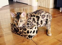
John K. Rosembert The Bengal cat is a domesticated cat breed created from hybrids of domestic cats, especially the spotted Egyptian Mau, with the Asian leopard cat, the breed name comes from the leopard cat’s taxonomic name. It is a long, muscular, medium- to large-sized cat, with a broad head and muzzle, high cheekbones, and pronounced whisker pads. The eyes are round and wide, with dark markings around the eyes (mascara) and the ears small and rounded at the tips. The grace of a jungle cat is held as one of the positive characteristics, along with the ability to move quietly and with stealth. The Bengal cat is known for its soft, sleek coat which has two main fur patterns: spotted (which is most common) and marbled. Both patterns are often tri-colored, giving each cat unique markings and patterns. This tri-coloring gives some Bengals spots which have a darker outline, often like the spots on a Jaguar. Because the Bengal is a hybrid developed from crossing the Asian Leopard cat and the American Shorthair. The result was a cat with an exotic look and a domestic temperament. The intelligent and athletic Bengal is an entertaining companion that demands human contact and can be very vocal in their pursuit of attention. The Bengal can be aggressive with other cats and needs to be socialized at an early age. They are highly active and agile, usually in constant motion. Bengals enjoy climbing, jumping, and a good game of fetch. Be sure to provide them with plenty of toys and a tall climbing tree to keep them amused and out of trouble. Below we will summarize some of the most major concerns of Bengal cat in order to help you prevent some predictable risks in your pet. Noted that before purchasing or adopting a Bengal, make sure the breeder offers a health guarantee on the kittens. Here are some of the most common diseases related to Bengal cat Joint problems: More often found in small dogs, Bengal cats may experience luxating patellas. luxating patella is a kneecap that slips off to the side of the leg because of an improperly developed stifle. A cat with a luxating patella may not show signs of pain or abnormality until the condition is well advanced; signs of this condition appear gradually and can progress to lameness as the cat grows older. Hip Dysplasia: another disease most commonly found in dogs, hip dysplasia may also occur in cats, especially in Bengals. Dysplasia is an inheritable condition that causes malformation of the hip joints and subsequent arthritis. Anesthetic Allergies: if your Bengal is going in for any type of surgery, including spaying and neutering, your vet must be careful regarding the use of anesthetics. Bengals, extremely sensitive to anesthetics, may experience allergic reactions that cause cardiac arrest. Always discuss the type of anesthetic with your vet before any surgery. Heart Disease: A heart condition, hypertrophic cardiomyopathy, is common in Bengals. This disease of the heart muscle usually occurs in the older cat. The heart muscle thickens, so the organ must work much harder, causing a number of problems. These may include blood clots, or thrombosis, rendering the back legs immobile. The disease also leads to congestive heart failure, resulting in death. Early signs of cardiomyopathy include panting and lethargy. https://www.petmd.com/cat/breeds/c_ct_bengal https://pets.thenest.com/bengal-cat-health-problems-4479.html https://animalhealthcenternh.com/client-resources/breed-info/bengal/ Photo credit: https://cattime.com/cat-breeds/bengal-cats
Feline Pancreatic Lipase
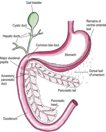
Feline Pancreatic Lipase LIN, WEN-YANG (WESLEY), Ph.D Feline Pancreatic Lipase is a powerful diagnosing biomarker for feline pancreatitis, whereas serum amylase and serum lipase are usually used in diagnosing pancreatitis for canine, are not effectively in detecting pancreatitis for cats. Anatomical physiology function of pancreas in cats The feline pancreas is a digestive glandular organ, which presents a long v-shaped strip of configuration locating at abdomen between stomach and duodenum. The tail part of feline pancreas rests toward the dorsal extremity of the spleen and connect to mesocolon with omentum (Figure 1). Figure 1. Anatomic view of the pancreas and its surrounding tissues https://veteriankey.com/pancreas-4/ The normal pancreas colored on pale pink and perform both functions of endocrine and exocrine. The endocrine portion contains small clusters of pancreatic α- and β-cells within Langerhans (approximately 2% of the gland’s weight) that majorly generates two hormones proteins: glucagon and insulin. Glucagon works to elevate the level of blood sugar, whereas insulin would diminish excess circulating blood sugar. Both of glucagon and insulin would take part in regulating homeostasis of glycemic level. Acinar and ductal cells are major members in the exocrine portion of pancreas. Acinar cells aggregate around the terminal pancreatic ductules to secrete digestive enzymes include trypsinogen, chymotrypsinogen, proelastase, procarboxypeptidase, ribonucleases, deoxyribonucleases, phospholipase A2, carboxylesterase amylase and lipase. Amylase and lipase are contributing to hydrolyze carbohydrates and fat. Others may play roles as proteases to cleave polypeptide chains. Feline’s ductal cell generates antibacterial proteins to protect small intestinal from bacterial infection. Moreover, ductal cell can also produce bicarbonate and water for neutralizing pH in the duodenum. Intrinsic factor such as vitamin B12 would be made from ductal cell too. The pancreas is the only generating organ of intrinsic factor for feline, whereas dogs could produce intrinsic factor from pancreas and stomach. All pancreatic enzymes would secret into small intestine for digesting fats, proteins and carbohydrates. Furthermore, the abnormal seep of exocrine enzymes to pancreas and its surrounding organs could cause pancreatic inflammation known as pancreatitis. The highest occurrence of feline pancreatic diseases is exocrine pancreatic insufficiency (EPI) and pancreatitis. Pancreatitis in the cat Feline pancreatitis is classified as acute (temporary morphological changes) and chronic (permanent morphological change) type according to the condition of histopathologic changes after treatment. Acute necrotizing pancreatitis (ANP) and acute suppurative pancreatitis are two of the most common types of acute feline pancreatitis, whereas chronic non-suppurative pancreatitis (CP) and pancreatic atrophy are chronic conditions. Common acute pancreatitis in feline would show anemia, leukocytosis, hypokalemia, hypocalcemia, hyperglycemia, elevation of ALT, ALP, total bilirubin, cholesterol and decreasing level of albumin. Acute necrotizing pancreatitis (ANP) usually present pancreatic acinar cell necrosis, peripancreatic fat necrosis followed inflammation, hemorrhage, mineralization and fibrosis. Despite of its idiopathic character, several diseases have considered to be related with development of ANP including concurrent biliary tract disease, ischemia, pancreatic ductal obstruction, toxoplasmosis, feline Herpes virus infections, feline infectious peritonitis, pancreatic fluke infestations (Eurytrema procyonis, Amphimerus pseudofelinus), trauma, organophosphate poisoning and hepatic lipidosis. General anesthesia induced hypotension or surgical venous outflow occlusion would decrease pancreatic blood flow and cause ANP. Acute suppurative pancreatitis is less common than ANP in feline and neutrophilic inflammation would happen with it. Besides, the continuous and progressive inflammatory process of the pancreas would lead to chronic non-suppurative pancreatitis (CP) in which lymphocytic inflammation, fibrosis, and acinar atrophy are the major features. The end stage of CP in most feline cases would usually result in pancreatic atrophy, which may or may not influence the endocrine portion of the gland. Felines who suffer pancreatic atrophy would occur cobalamin and fat-soluble vitamin malabsorption, severe maldigestion, acid injury in duodenal mucosa, and bacterial proliferation in the gut due to exocrine pancreatic insufficiency. Since various types of pancreatitis such as acute and chronic pancreatitis, pancreatic abscess, pancreatic cyst/pseudocyst, exocrine pancreatic insufficiency, and neoplasia share overlapping symptoms, thus histopathology cab be used to discern different conditions. Symptoms of feline pancreatitis Symptoms of feline pancreatitis would occur lethargy, anorexia, dehydration, hypothermia, vomiting, weight loss, Jaundice, cholangiohepatitis, hepatic lipidosis, biliary obstructions, cranial abdominal masses and cranial abdominal discomfort. Besides, hepatic and intestinal disease would concurrent. I. Acute feline pancreatitis: Vomiting, poor appetite, poor activity, diarrhea, abdominal pain, drooling, fever, collapse. II. Chronic feline pancreatitis: Frequent vomiting, poor appetite, listless, frequent diarrhea, abdominal pain, drooling, fever, collapse, hypothermia, breathing too fast or too slow, fast heartbeat. Possible Causes of feline pancreatitis Scientists presumed that premature trypsin activate of digestive zymogens in pancreatic acinar cells would cause pancreatic autodigestion, acinar cell necrosis, hemorrhage, and fat necrosis, saponification, mast cell degranulation, leukocyte chemotaxis, platelet aggregation, vasodilation, surfactant degradation within the lungs and initiation of disseminated intravascular coagulation (DIC) for worse cases. Diagnosis and general considerations Since symptoms of feline pancreatitis were similar to common flu, vets probably can’t judge it correctly at the early stage without proper diagnostic tools. It’s not an easy task to diagnose pancreatic disease within cats. Single diagnostic method is not recommended. The diagnosis of feline pancreatitis relied on combinational diagnostic tools include historical data, physical examination results, laboratory, and Bio-imaging. Especially, histopathology plays the role as the definitive conclusion for feline pancreatitis. Middle-aged felines are susceptible to pancreatitis; whereas, older felines (mean 12.8 years, range 4–20 years) are susceptible to neoplasia, cyst/pseudocysts. Diagnostic imaging Radiography, computed tomography (CT) and ultrasonography were effective imaging tools applied in diagnosing feline pancreatitis. Among all imaging tools, ultrasonography is the most reliable imaging modality for the diagnosis of feline pancreatic diseases, which can help identify soft tissue masses, cysts/pseudocysts, abscesses, or neoplasia and lesions of feline pancreatitis. Clinicopathologic tests The general clinicopathologic test for feline pancreatitis include complete blood cell count, serum bilirubin, cholesterol, glucose, total protein, albumin, and serum activity of liver enzymes (serum alanine aminotransferase and alkaline phosphatase), calcium, urea, creatinine, and potassium. However, serum lipase and amylase activities can’t be diagnostic indicator for feline pancreatic disease. Nevertheless, feline trypsin-like immunoreactivity (fTLI) and