
Breed-related disease: Dalmatians
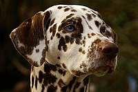
John K. Rosembert Dalmatians are one of the oldest dog breeds known to man, although their exact origins are somewhat shrouded. The earliest recorded history of the breed places them in regions of Asia and Europe, particularly in Dalmatia, and it’s from here that the breed takes its name. Historically, Dalmatians have been known as coaching dogs, running around and beneath carriages, following their master’s travels. They are also known as the fireman’s friend, and once traveled along on runs in the days of the horse-pulled fire wagon. Dalmatians are a large, strong, muscular dog. The skull is about as wide as it is long, and flat on the top. The muzzle is about the same length as the top of the skull. The stop is moderate but well defined. The nose can be black, brown (liver), blue or a dark gray that looks like black. The teeth meet in a scissors bite. The medium-sized round eyes are brown, blue or a combination of both. The ears are set high, hanging down, gradually tapering to a rounded tip. The chest is deep. The base of the tail is level with the topline and tapers to the tip. The feet are round with arched toes. Toenails are white and/or black in black-spotted dogs and brown and/or white in liver-spotted dogs. The short coat has fine dense hairs. The symmetrical coat is predominantly white with clearly defined round spots. The spots can be black or brown (liver) which are the preferred colors in the show ring, but can also be, lemon, dark blue, tricolored, brindled, solid white or sable. Not all of these colors are accepted into the show ring, but they do occur in the breed. The more defined and well distributed the markings are, the more valued the dog is to the show ring. Puppies are born completely white and the spots develop later. They are highly energetic, playful and sensitive dogs. They are loyal to their family and good with children, although some Dalmatian experts caution that the breed may be too energetic for very small children. These dogs are intelligent, can be well trained and make good watchdogs. Some Dalmatians can be reserved with strangers and aggressive toward other dogs; others are timid if they are not well socialized, and yet others can be high-strung. These dogs are known for having especially good “memories” and are said to recall any mistreatment for years. Like other breeds, Dalmatians display a propensity towards certain health problems specific to their breed, Such as: Deafness A genetic predisposition for deafness is a serious health problem for Dalmatians; American Dalmatians exhibit a prevalence for bilateral congenital sensoneural deafness of 8%, (for which there is no possible treatment), compared with 5.3% for the UK population. Deafness was not recognized by early breeders, so the breed was thought to be unintelligent. Many breeders, when hearing testing started to become the norm, were amazed to discover that they had uni hearing dogs. Even after recognizing the problem as a genetic fault, breeders did not understand the dogs’ nature, and deafness in Dalmatians continues to be a frequent problem. Dalmatian-Pointer Backcross Project Hyperuricemia in Dalmatians (as in all breeds) is inherited, but unlike other breeds, the “normal” gene for a uric acid transporter that allows for uric acid to enter liver cells and be subsequently broken down is not present in the breed’s gene pool. Therefore, there is no possibility of eliminating hyperuricemia among pure-bred Dalmatians. The only possible solution to this problem must then be crossing Dalmatians with other breeds to reintroduce the “normal” uric acid transporter gene. This led to the foundation of the Dalmatian-Pointer Backcross Project, which aims to reintroduce the normal uric acid transporter gene into the Dalmatian breed. Hyperuricemia Dalmatians, like humans, can suffer from hyperuricemia, Dalmatians’ livers have trouble breaking down uric acid, which can build up in the blood serum (hyperuricemia) causing gout. Uric acid can also be excreted in high concentration into the urine, causing kidney stones and bladder stones. These conditions are most likely to occur in middle-aged males. Males over 10 are prone to kidney stones and should have their calcium intake reduced or be given preventive medication. Eye Problems Not many things have as dramatic an impact on your dog’s quality of life as the proper functioning of his eyes. Unfortunately, Dalmatians can inherit or develop a number of different eye conditions, some of which may cause blindness if not treated right away, and most of which can be extremely painful!. Sources: https://en.wikipedia.org/wiki/Dalmatian_(dog) https://www.dogbreedinfo.com/dalmatian.htm Photo credit: https://en.wikipedia.org/wiki/Dalmatian_(dog)
Breed-related disease: Burmilla

The Burmilla started out as an accident. In 1981, a Chinchilla Persian male and a Lilac Burmese bred, and the female delivered four kittens. These kittens had an unusual black-tipped coloring. The look of these cats was so attractive that a breeding program was inaugurated to produce a cat that would have the short hair of the Burmese, the roundness taken from both breeds, and the unusual coloring seen in the initial kittens. The Burmilla is rarely seen. In Britain, it is still an experimental breed, and it is not yet accepted by the major registries in the United States. The Burmilla is a medium-sized cat, but she is also stocky and heavy. This breed is somewhat compact while being very muscular with heavy boning. It is a cat that is very rounded. The head is round and the tips of the ears are round. The profile shows a “break,” and the eyes are very slightly slanted. The coat of the Burmilla is short and soft. Because of the original pairing, the coat is also thick and dense. The Burmilla is a fairly placid cat. She tends to be an easy cat to get along with, requiring minimal care. The Burmilla is affectionate and sweet and makes a good companion. They are good climbers and jumpers and should have cat trees and perches. The Burmilla is a sturdy, stocky cat and you might have to watch her weight carefully, particularly if she does not get enough exercise. Modify her nutrition if you need to do so. Burmilla’s are a typically healthy breed with an expected lifespan of 7–12 years. Therefore they are also prone to a few health issues, Such as: Congenital keratoconjunctivitis sicca causes dry eyes, chronic conjunctivitis and corneal vascularization Feline Orofacial pain syndrome affects male cats in particular, although females are also affected. Symptoms include exaggerated licking and chewing movements, plus excessive pawing at the mouth. This happens in distinct episodes, although the cat remains alert (albeit in distress) for the duration . The disease appears to be related to some kind of oral pain or distress, and is possibly linked to teething or dental disease. A possible risk factor is stress, but it is believed that there are also hereditary factors involved. Polycystic Kidney Disease (PKD ) an inherited kidney disease where cysts form in the kidneys at birth, gradually increasing in size as the cat ages. PKD eventually leads to kidney failure, however, it can be managed to help decrease the workload on the kidneys. Sources: https://www.petinsurance.com/healthzone/pet-breeds/cat-breeds/burmilla/ https://www.dailypaws.com/cats-kittens/cat-breeds/burmilla# : Photo credit: https://www.worldlifeexpectancy.com/cat-life-expectancy-burmilla
Breed-related disease: Basset Hound

Basset Hounds were originally bred in France and Belgium (“basset” is French for “low”). It is thought that the friars of the Abbey of St. Hubert were responsible for crossing strains of older French breeds to create a low-built scenting hound that could plod over rough terrain while followed on foot by a human hunting partner tracking rabbit and deer. Their accuracy and persistence on scent made Bassets a popular choice for French aristocrats, for whom hunting was a way of life. Bassets are very heavy-boned dogs with a large body on fairly short legs. Because they are bulky, bassets are slow maturing dogs, often not reaching full size until two years old. Bassets are immediately recognizable by their short, crooked legs, their long hanging ears and their large heads with hanging lips, sad expressive eyes, and wrinkled foreheads. The tail curves up and is carried somewhat gaily. The body is long and with the short legs gives bassets a rectangular appearance. The basset has a nice short, tight coat, with no long hair on legs or tail. Colors most commonly seen are tricolor or red and white but any hound color is acceptable. The Basset Hound is among the most good natured and easygoing of breeds. This breed is amiable with dogs, other pets, and children, although children must be cautioned not to put strain on this and all dogs’ backs with their games. The Basset is calm inside, but needs regular exercise in order to keep fit. They prefer to investigate slowly, and love to sniff and trail. These are talented and determined trackers, not easily dissuaded from their course. Because of this, they may get on a trail and follow it until becoming lost. This dog tends to be stubborn and slow moving. Bassets have a loud bay that they use when excited on the trail. Here, we summarized the most common health concerns you should look for over the lifetime of your Basset. Here we go: Bloat Gastric Dilatation and Volvulus, also known as GDV or Bloat, usually occurs in dogs with deep, narrow chests. This means your Basset is more at risk than other breeds. When a dog bloats, the stomach twists on itself and fills with gas. The twisting cuts off blood supply to the stomach, and sometimes the spleen. Left untreated, the disease is quickly fatal, sometimes in as little as 30 minutes. Your dog may retch or heave (but little or nothing comes out), act restless, have an enlarged abdomen, or lie in a prayer position (front feet down, rear end up). Back Problems Intervertebral disc disease (IVDD) is a common condition in dogs with long backs and short legs, which may include your Basset. The disease is caused when the jelly-like cushion between one or more vertebrae slips or ruptures, causing the disc to press on the spinal cord. If your dog is suddenly unable or unwilling to jump up or go upstairs, is reluctant to move around, has a hunched back, cries out, or refuses to eat or go potty, he is likely in severe pain. He may even drag his back feet or be suddenly paralyzed and unable to get up or use his back legs. If you see symptoms, don’t wait. Call us or an emergency clinic immediately! For less severe cases, rest and medication may resolve the problem. Joint Disease When Basset puppies are allowed to grow too quickly, the cartilage in their joints may not attach to the bone properly. This problem is known as osteochondritis dissecans or OCD. If this occurs, surgery may be required to fix the problem. It’s best to stick to our recommended growth rate of no more than four pounds per week. Don’t overfeed him and don’t supplement with additional calcium. Feed a large-breed puppy diet rather than an adult or a regular puppy diet. Bleeding Tumor Hemangiosarcoma is a type of bleeding tumor that affects Basset Hounds at greater than average incidence. These tumors commonly form in the spleen, but can form in other organs as well. Unbeknownst to a pet owner, the tumor breaks open and internal bleeding occurs. Some tumors can be volleyball-sized or larger before signs of sickness show. We often find clues that one of these tumors is present during senior wellness testing, so have his blood tested and an ultrasound performed at least yearly. Sources: https://www.petfinder.com/dog-breeds/basset-hound/ https://coonrapidspethospital.com/client-resources/breed-info/basset-hound/ Photo credit: https://mom.com/momlife/19480-cool-facts-about-basset-hounds/
Pneumonia and gastritis in a cat caused by feline herpesvirus-1
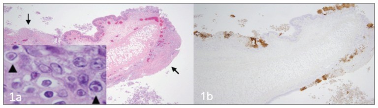
Source: https://www.ncbi.nlm.nih.gov/pmc/articles/PMC4712990/ A fatal respiratory and gastric herpesvirus infection in a vaccinated, 6-year-old neutered male domestic shorthair cat with no known immunosuppression or debilitation. Histology examination revealed severe necrotizing bronchopneumonia, fibrinonecrotic laryngotracheitis, and multifocal necrotizing gastritis associated with eosinophilic intranuclear inclusion bodies in affected tissues of larynx, trachea, lung and stomach. Immunohistochemistry also displayed strong immunoreactivity for FHV-1 in the corresponding section of larynx, trachea, lung and stomach. Figure 1 Larynx. Cat. a — Note the multifocal areas of ulceration (arrows) and inflammation. Inset: inclusion bodies (arrowheads) within epithelial cells adjacent to areas of ulceration. H&E. b — Immunohistochemistry of the corresponding section of larynx displaying strong multifocal immunoreactivity for FHV-1. Figure 2 Trachea. Cat. a — Note the denuded tracheal mucosa covered by a thick fibrinonecrotic exudate (asterisks), attenuation of the epithelium lining the tracheal glands (arrows) and numerous inflammatory cells infiltrating the tracheal wall. Inset: Inclusion bodies within the epithelial cells lining the tracheal glands (arrowheads). H&E. b — Strong immunoreactivity for FHV-1 in the epithelium of the trachea and tracheal glands. Figure 3 Lung. Cat. a — Severe neutrophilic necrotizing bronchopneumonia affecting airways (asterisks) and surrounding tissues. H&E. b — Strong immunoreactivity for FHV-1 in bronchial and bronchiolar epithelium. Figure 4 Stomach. Cat. a — Area of necrosis within the gastric mucosa (asterisk). H&E. b — Multifocal areas of FHV-1 immunoreactivity corresponding to areas of necrosis within the gastric mucosa.
Feline Tritrichomonas Foetus Infection: A Review
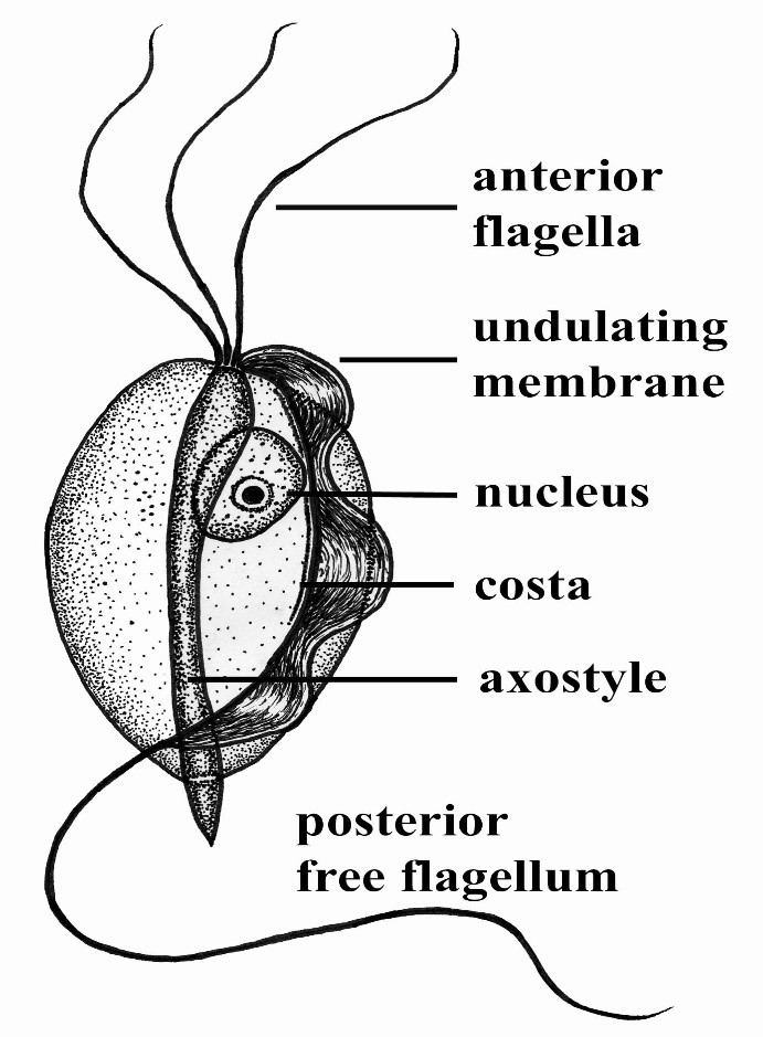
Maigan Espinili Maruquin Structure and Epidemiology The protozoan parasite Tritrichomonas foetus has sudden emergence of its syndrome in the 1990s causing feline intestinal tritrichomoniasis and has then attracted feline medicine studies (Levy, Gookin et al. 2003, Bell, Gowan et al. 2010) (Levy, Gookin et al. 2001 ). At a range of 10- 31%, infected cats in UK and USA comes from young, pedigree cats in multi-cat environment (Gookin, Levy et al. 2001, Foster, Gookin et al. 2004, Gookin, Stebbins et al . 2004, Gunn-Moore, McCann et al. 2007, Frey, Schild et al. 2009, Stockdale, Givens et al. 2009)( Gunn-Moore D, Tennant B, 2007)(Bell, Gowan et al. 2010). An estimate 30% of purebred cats in USA are suggested infected by T. foetus (Gookin, Stebbins et al. 2004, Gookin, Stauffer et al. 2010). The T. foetus , like other trichomonads, has only trophozoite stage, however, it has a pseudocyst stage (Lipman NS, et. al., 1999) (Pereira-Neves, Ribeiro et al. 2003, Benchimol 2004, Mariante, Lopes et al. 2004, Yao and Köster 2015). Therefore, cats are presumed to be infected via direct contact due to the absence of the cyst stage (Zajac AM, Conboy GA, 2006). The parasite is pear- or spindle-shaped, having three anterior flagella and one posterior flagellum. With its size of approximately 10-25 μm in length and 3-15 μm in width, the undulating membrane extends along the whole length of the body which emerges as the posterior flagellum. This trophozoite reproduces asexually by longitudinal binary fission (Yao and Köster 2015). (https://www.k-state.edu/parasitology/625tutorials/Protozoa10.html) Fig. 01. Morphologic structure of the Tritrichomonas foetus Infected cats were reported to experience chronic large bowel diarrhea as the parasite was found in the feline intestine (Foster, Gookin et al. 2004, Payne and Artzer 2009). Reports also showed the presence of the organism in the feline uterus (Dahlgren, Gjerde et al. 2007). Further, the isolates of T. foetus from cattle are known to be infectious for the cats, while the isolates of the same species from cats are infectious to cattle (Stockdale, Dillon et al. 2008, Walden, Rodning et al. 2008, Payne and Artzer 2009). Clinical Signs/ Pathogenesis Despite suggestions of strong association of feline T. foetus and chronic diarrhea (Gookin, Stebbins et al. 2004, Mardell and Sparkes 2006, Gunn-Moore, McCann et al. 2007, Burgener, Frey et al. 2009, Holliday, Deni et al. 2009, Pham 2009, Stockdale, Givens et al. 2009, Kuehner, Marks et al. 2011), whether the parasite alone is sufficient to cause clinical signs or the foetus-associated diarrhea, being a primarily multifactorial disease involves concurrent infection with other enteropathogens, host and environmental factors (Gookin, JL, et. al., 1999) (Gookin, Levy et al. 2001, Bissett, Gowan et al. 2008, Stockdale, Givens et al. 2009, Kuehner, Marks et al. 2011). It is possible that the trophozoites are transmitted by a fecal- oral route from an infected to uninfected cat (Yao and Köster 2015). Symptoms may appear early as 2 to 7 days after orogastric inoculation (Gookin, Levy et al. 2001, Yao and Köster 2015). Whereas, infected cats showed anorexia, depression, vomiting and weight loss while experimental infections also reported vomiting and fever (Mardell and Sparkes 2006, Xenoulis, Lopinski et al. 2013, Yao and Köster 2015)( Stockdale, H., et al., 2007). Chronic large bowel diarrhea, associated with blood, mucus, flatulence, tenesmus, and anal irritation were also reported (Foster, Gookin et al. 2004, Gookin, Stebbins et al. 2004, Payne and Artzer 2009, Stockdale, Givens et al. 2009). With the characteristic of the trichomonads as commensal organisms, some hosts show no clinical signs and are asymptomatic (Payne and Artzer 2009). In some studies, the parasite, with its surface located antigen, was detected on epithelial surface and within the superficial detritus of the cecal and colonic mucosa (Gookin, Levy et al. 2001, Yao and Köster 2015), while naturally infected cats showed parasite in close proximity to the mucosal surface and less frequently in the lumen of colonic crypts (Yaeger and Gookin 2005, Yao and Köster 2015). Conclusively, T. foetus trophozoites can be detected in epithelial surface and crypts of cecum and colon (Yao and Köster 2015). Mechanisms were then described to include possibilities of alterations in the normal intestinal flora, adherence to the epithelium, and elaboration of cytokines and enzymes (Payne and Artzer 2009). Diagnosis For cats <6months old with recent clinical signs of chronic large bowel diarrhea, infection with T. foetus infection is suspected (Yao and Köster 2015). Diagnosis of the T. foetus infection may be conducted via direct observation of the flagellates in fresh or cultured feces (Payne and Artzer 2009) or on a saline diluted direct fecal smear (Yao and Köster 2015). The trophozoites of T. foetus are difficult to distinguish from Giardia spp and other nonpathogenic intestinal trichomonads. However, although the size of T. foetus and Giardia spp are almost the same, they move differently. The movement of Giardia spp. resembles the fall of a leaf while trichomonads move erratically (Yao and Köster 2015)( Gookin JL, Levy MG, 2008). Feline feces can be cultivated and be tested in commercially available InPouch™ TF medium (Payne and Artzer 2009, Yao and Köster 2015) or DNA extraction and amplification of T. foetus rDNA by the use of PCR from feces samples can be conducted (Gookin, Stebbins et al. 2004, Manning 2010, Yao and Köster 2015). Moreover, other causes of diarrhea, like bacterial, viral, other parasites, and nutritional problems should be ruled out before a diagnosis of tritrichomoniasis can be made (Payne and Artzer 2009). Treatment and Disease Managements The T. foetus infection in cats has no approved treatment. While treating infected animals is difficult, success is also limited (Payne and Artzer 2009). Literatures have used therapeutics including paromomycin, fenbendazole, furazolidone, nitazoxanide, metronidazole, tinidazole and ronidazole (Gookin, Breitschwerdt et al. 1999, Gookin, Levy et al. 2001, Yao and Köster 2015). The ronidazole is not registered for human or veterinary use (Yao and Köster 2015) however, it showed effectiveness in experimentally infected cats (Gookin, Copple et al. 2006) (Gookin JL, Dybas D, 2008). Due to possible neurologic side effects, caution is observed
Leptospirosis
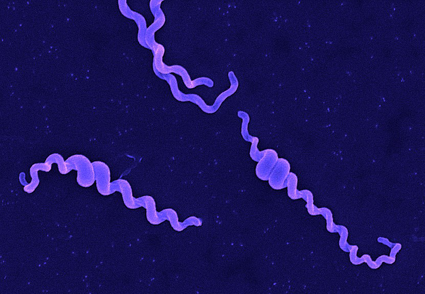
Andy Hua Introduction Classification Taxonomy Classically, the genus Leptospira was divided into 2 species based on genetic analysis: L. interrogans sensu latu (pathogenic strains) and L. biflexa sensu latu (saprophytic strains). L. interrogans is divided into more than 250 serovars based on antigenic composition and further classified into antigenically related serogroups. There is a serovar spectrum and frequency that differs according to countries and regions (depending on distribution of rodent hosts, import of dogs from abroad, use of vaccination). The main infecting serovars in dogs were Icterohaemorrhagiae and Canicola in Europe and America prior to 1960. Since the use of the bivalent vaccine against Canicola and icterohaemorrhagiae , OTHER serovars A to the Shift occurred .Besides. L. icterohaemorrhagiae and L. canicola , serovars Importance of Dogs in the include: grippotyphosa , Bratislava , Saxkoebing , Sejroe , Copenhagi , Australis , Bataviae , and Pomona , autumnalis , and hardjo . Icterohaemorrhagiae and Canicola infections in unvaccinated dogs still occur, indicating that these serovars are not fully eradicated. Leptospires are motile, obligate aerobe, gram-negative bacteria, which are not visible in routinely fixed smears. Dark field microscopy (Fig. 1) or phase contrast microscopy is necessary for visibility of unstained leptospires. Clinical Effects Epidemiology Habitat Leptospires have been isolated from birds, reptiles, amphibians and invertebrates. Rodents and wild carnivores are the most frequent carriers. Reservoir hosts show few or no signs of disease. Leptospira spp. are commonly sequestered in the renal tubules of mammalian kidneys. Different serovars typically have different reservoir hosts. Lifecycle Generation time in culture media or host is long. Transmission Direct or indirect transmissions are possible. Indirect transmission through contaminated water or soil is more common. Pathological effects Infection occurs through ingestion of infected rodents or penetration of mucosae or traumatized skin. Leptospiremia occurs within 1 week. Leptospires spread to other organ systems (kidneys, liver, spleen, endothelial cells, lungs, uvea/retina, skeletal and heart muscles, pancreas, and genital tract) and cause tissue damage, visceral and vascular inflammation. Leptospiral pulmonary hemorrhage syndrome (LPHS) Lung: pulmonary hemorrhage can occur as severe manifestation of acute leptospirosis. Leptospires can persist in immune privileged site (eg, renal tubes, eye). In the presence of adequate antibody titers, leptospires are eliminated from most organs. In the presence of low antibody titers mild leptospiremia can continue with a subclinical course of disease. Other Host Effects Individual host may show little or no clinical signs but may be source of infection in the same animal species. An animal that has recovered may become a long-term shedder of the organism. Mainly dogs show disease, rodents often the reservoir. Cat disease is uncommon, but serology shows that asymptomatic infection occurs. Individual host can show little or no clinical signs but can be source of infection to other animals or humans. An animal that has recovered can become a long-term shedder of the organism. Rodents are often the reservoir. Dogs commonly succumb to disease if infected. In cats, disease is uncommon but asymptomatic infection and shedding in urine occurs. Control Control via animal Antimicrobial therapy Dogs with gastrointestinal signs should initially be treated with intravenous penicillin derivates (eg, ampicillin or amoxicillin 20-30 mg/kg q6-8h). These should be continued until gastrointestinal signs are under control and liver enzymes are normalized. A directly following antimicrobial therapy with 3 weeks of oral doxycycline (5 mg/kg q12h) is necessary for prevention of carrier states. Dogs without gastrointestinal signs should immediately be treated with doxycycline. Antibody testing of dogs living in the same household as infected dogs is recommended. Oral doxycycline (5 mg/kg q12h for 3 weeks) should be administered, if these dogs have antibodies. Symptomatic treatment Treatment of dogs with gastrointestinal sings includes antiemetics, gastroprotectants, and nutritional support. Use of opioids in dogs with pain can be necessary. Treatment of dogs with acute kidney injury (AKI) includes correction of loss of fluid, electrolytes, acid-base imbalances and hypertension, and if necessary hemodialysis for patients with persistent oligoanuria, life-threatening hyperkalemia, or severe volume overload. Oxygen therapy or mechanical ventilation can be necessary in dogs with LPHS. Plasma transfusions can be necessary for patients with DIC (disseminated intravascular coagulation). Whole blood transfusion can be helpful, if bleeding occurs. Hemodialysis Hemodialysis is necessary in dogs with acute renal failure (life-threatening hyperkalemia or severe volume overload) and in dogs with advanced uremia refractory to medical management. Early referral to facilities where hemodialysis is available is recommended. Renal recovery usually occurs after 2-7 days of dialytic support. Hemodialysis leads to favourable prognosis for renal recovery (in more than 80% of dogs). Mechanical ventilation Anesthesia ventilators: overview can be necessary in dogs with severe pulmonary hemorrhage due to LPHS. Vaccination Vaccination protects against clinical disease and carrier status with shedding. Protection is serogroup-specific and temporary. Annual boosters are required. Diagnosis Leptospirosis at its onset is often misdiagnosed as aseptic meningitis, influenza, hepatic disease or fever (pyrexia) of unknown origin. Despite being common, the diagnosis of leptospirosis is often not made unless a patient presents with textbook manifestations of the so called Weil’s disease, such as fever plus jaundice, renal failure and pulmonary haemorrhage. Leptospiral infection often has minimal or no clinical manifestations; of the cases in which fever develops, as many as 90% are undifferentiated febrile illnesses. Moreover, clinicians may fail to recognize that transmission of leptospirosis can occur in the urban setting because it is incorrectly perceived to be a rural disease. Therefore, diagnosis is based on laboratory tests rather than on clinical symptoms alone. In developing countries,Laboratory facilities may be inadequate for diagnosis a high prevalence of the disease. Of substantial clinical importance despite the syndrome of leptospiral pulmonary haemorrhage has emerged in recent years, in diverse places around the world. Two important issues continue to confront clinicians regarding leptospirosis. The first is how to reliably establish the diagnosis. The most common way to diagnose leptospirosis is through serological tests either the Microscopic Agglutination Test (MAT) which detects serovar-specific antibodies, or a solid- phase assay for the detection of Immunoglobulin M (IgM) antibodies. Leptospira are present in the blood until they are
Breed-related disease: Cymric cat
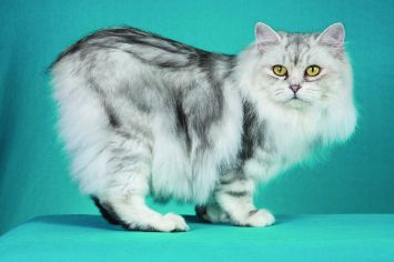
Is it really a cat if it doesn’t have a tail? It is if it’s a Cymric (pronounced kim-rick). There are lots of cats with short tails or no tails, but the Cymric (and his sister breed the shorthaired Manx) is the only one specifically bred to be tail-free. Sometimes jokingly said to be the offspring of a cat and a rabbit (however cute the idea, a “cabbit” is biologically impossible), these particular tailless cats are the result of a natural genetic mutation that was then intensified by their remote location on the Isle of Man, off the coast of Britain. The cats are thought to date to 1750 or later, but whether a tailless cat was born there or arrived on a ship and then spread its genes throughout the island cat population is unknown. The island became known for tailless cats, and that is how the breed got its name of Manx. The Manx has long been recognized by the Cat Fanciers Association, The International Cat Association, and other cat registries. A longhaired version was accepted by CFA as a division of the Manx in 1994. In some associations, the longhaired Manx is called a Cymric and is considered a separate breed. The overall appearance should be that of a medium-sized, compact, muscular cat. The Cymric has a round head with a firm muzzle and prominent cheeks, short front legs, height of hindquarters, great depth of flank, and a short back, which forms a smooth continuous arch from the shoulders to the round rump. The Manx and Cymric are essentially the same in all respects, the Cymric having a longer coat. The Cymric has a medium/semi-long coat with a silky texture, which varies with coat color. Britches, tufts of hair between the toes and full furnishings in the ears distinguish the Cymric. The personality of the Cymric has won a strong following. Cymrics are intelligent, fun-loving cats, and they get along well with other pets, including dogs. Cymrics are particularly noted for their loyalty to their humans and enjoy spending quality time with them. As cats go, they can be easily taught tricks. Despite their playful temperament, they are gentle and nonaggressive. Here below, we listed some of the most common diseases to look for in your Cymric: Congenital Vertebral Malformations Sacrocaudal dysgenesis is a form of spinal deformity commonly seen in Cymric kittens. The sacrum is the part of the spine that passes through the pelvis, caudal means “towards the tail”, and dysgenesis means improper formation during fetal development. Sacrocaudal dysgenesis, then, means that the tail end of the spine forms improperly during fetal development—a common problem when a cat’s tail is genetically programmed to be missing. Affected kittens whose spine and spinal nerves aren’t functioning correctly may have fecal or urinary incontinence, or may walk with a hopping, abnormal gait. Constipation and megacolon are also more common in affected cats, and the effects of the condition typically worsen with age. Megacolon Megacolon is a serious and chronic form of constipation caused by a defect in the nerve supply to the intestines. Cats with megacolon have an insufficient number of nerves linked to the muscles of the colon and are unable to pass stool properly. As the cat gets older, the condition worsens, causing an increase in discomfort and blockage. Each time the cat is constipated, the intestines are stretched, bruised, and further damaged. This damage leads to larger and harder stools and, eventually, complete blockage. Megacolon is a life-threatening condition, but early and aggressive treatment can delay or prevent the necessity of major surgery to remove the colon. FLUTD When your cat urinates outside the litter box, you may be annoyed or furious, especially if your best pair of shoes was the location chosen for the act. But don’t get mad too quickly—in the majority of cases, cats who urinate around the house are sending signals for help. Although true urinary incontinence, the inability to control the bladder muscles, is rare in cats and is usually due to improper nerve function from a spinal defect, most of the time, a cat that is urinating in “naughty” locations is having a problem and is trying to get you to notice. What was once considered to be one urinary syndrome has turned out to be several over years of research, but current terminology gathers these different diseases together under the label of Feline Lower Urinary Tract Diseases, or FLUTD. Sources: https://ahcfargo.com/client-resources/breed-info/cymric-longhaired-manx-2/ https://cattime.com/cat-breeds/cymric-cats#/slide/1 Photo credit: https://en.wikipedia.org/wiki/Cymric_cat https://www.petfinder.com/cat-breeds/cymric/
Breed-related disease: Maltese dog

John K. Rosembert The Maltese is an ancient breed, one of several small “bichon” dogs found around the Mediterranean for thousands of years. His exact place of origin is a mystery, with conjecture including Sicily, Egypt, and southern Europe, but most historians pinpoint Malta for the development of the breed. With its teeny-tiny stature, flowing white coat, and high trainability, this toy breed has beauty and brains. For that, it’s been cherished since its earliest days in ancient Italy and has long been seen as a portable and charming companion. The Maltese puppy truly is the quintessential lap dog, with its fluffy white fur, adorable black-button nose, dark eyes, and sprightly demeanor. “They’re like a little stuffed animal,” Derse says. The Maltese has a compact, athletic body, small floppy ears, and a tufted tail that curves over her back. By the time a Maltese reaches her full 7–9 inch height and 4–6 pound weight, those white tresses become silky smooth, requiring daily brushing along with regular baths to maintain their regal appearance. If you’re looking for a friendly dog with elegance and charm, look no further than the Maltese! With their silky, pure white coats and warm temperaments, it’s no surprise that they’ve made popular companions for people for centuries. People fall head over heels for this breed because of its adorable look, but their loyal personality makes them a fantastic option for anyone that’s looking for a canine friend. You will find no lack of adorableness, loyalty, or intelligence in this famous toy dog breed. Like other toy breeds, Maltese are prone to certain health issues. However, some of these conditions can be prevented if you keep up with routine care and checkups, here below we listed some of the most common illnesses run in their genes… Let’s get started: Inflammatory Bowel Disease Other common health conditions Maltese face affect their intestines. The breed can also develop severe food allergies and sensitivities. Inflammatory Bowel Disease, or IBD for short, occurs when your dog’s intestines become overactive with lymphocytes and plasmacytes. These are immune cells that attack harmful organisms in your dog’s gut, but they can cause vomiting and diarrhea when they become overactive. Diet and lifestyle changes will help this condition. Epilepsy Seizures and epilepsy can occur in Maltese as well. These conditions tend to be genetic, so if the dog’s parents or other direct relatives have a history of seizure, then your dog will have a higher chance of experiencing them. Because Maltese dogs have low body weight, they are at risk of experiencing hypoglycemic seizures. Hypoglycemic seizures are caused by low blood sugar levels. Seizures start as an excessive surge of electrical activity in the brain. The surge becomes excessive and overwhelms the neurons, resulting in a temporary malfunction of the brain. Depending on which regions of the brain are affected, a seizure manifests as muscle spasms or other symptoms. Knee Problems Sometimes your Maltese’s kneecap (patella) may slip out of place (called patellar luxation). You might notice that he runs along and suddenly picks up a back leg and skips or hops for a few strides. Then he kicks his leg out sideways to pop the kneecap back in place, and he’s fine again. If the problem is mild and involves only one leg, your friend may not require much treatment beyond arthritis medication. When symptoms are severe , surgery may be needed to realign the kneecap to keep it from popping out of place. Heart murmurs, congestive heart failure This is most often seen with senior Maltese dogs age 10 and up. Because heart murmurs rarely have any outward signs, this is usually discovered during a wellness check when the veterinarian is listening to the dog’s heart. Murmurs do not always lead to congestive heart failure, but they can. These are graded on a scale from 1 to 6. Typically no treatment is required for a grade 1 to 3 murmur. However, this is often a progressive disease. If the murmur worsens to a grade 4, 5, or 6, there can be issues such as troubled breathing, coughing, and exercise intolerance. Sources: https://www.dailypaws.com/dogs-puppies/dog-breeds/maltese https://www.petmaltese.com/maltese-health https://www.holistapet.com/maltese-dog-breed-temperament-personality/ Photo credit: https://thehappypuppysite.com/maltese-lifespan/ https://maltaangelmaltese.com/about-us/
Breed-related disease: Birman Cat

The origin of the Birman cat is not well known, with much of his history tied in with cultural legends, While there is no clear record of the origin of Birman cats, one pair was taken to France around 1919, from which the breed became established in the western world. However, Birman cats were almost wiped out as a breed during World War II and were heavily outcrossed with long-hair breeds (mainly Persians) and also Siamese lines to rebuild the breed. By the early 1950s, pure Birman cat litters were once again being produced. The restored breed was recognized in Britain in 1965. The Birman, also known as the “Sacred Cat of Burma” is semi-longhaired with darker coloring to the points, face, legs, ears, and tail, and a pale toning body color. It is a largish cat with a thickset body and short legs.The Birman cat has blue eyes and four pure white feet. The front gloves covering only the feet, but the rear socks are longer. The head is broad and rounded with medium-size ears. They come in lots of different colors. The Birman is a calm, affectionate cat who loves to be around people and can adapt to any type of home. He likes to play chase with other pets, taking turns being the chaser and the one being pursued. Birmans make friends with kids, dogs , and other cats. In fact, unlike most felines, they don’t especially like being the “only pet,” so you may want to get your Birman a companion – he won’t care if it’s another Birman, a different breed of cat, or even a dog. Birmans aren’t demanding of your attention, but they’ll definitely let you know when they need a head scratch or some petting. Then they’ll go about their business until it’s time for you to adore them again. You should also keep your Birman entertained with interactive toys that require him to do some thinking and moving to pop out treats or kibble. Here we gathered for you the most common Genetic Predispositions about Birmans, let’s get started: Luckily, Birman cats are relatively healthy and aren’t predisposed to any major conditions. But, the common health concerns that plague other cats are things to look out for. These include obesity, hypertrophic cardiomyopathy, and kidney disease. Obesity Because of the hefty weight of these kitties, they may be more prone to feline obesity, which can cause a myriad of other health concerns. Just like humans, it’s important to do everything you can to help your kitty maintain a healthy weight. By limiting their food intake, exercising them regularly, and keeping up with regular vet visits, you can completely prevent this condition. It is up to the owner of Birmans to make sure they stay at a healthy weight. Hypertrophic Cardiomyopathy (HCM) Not specific to Birman cats, hypertrophic cardiomyopathy (HCM) is the most common heart condition in cats. It causes the walls of the heart muscle to thicken and can cause the heart to increase in size. This is a genetically inherited condition, and breeders can check their lines for this condition. HCM is something to always be aware of, even if your kitty has a clean bill of health. HCM ranges in severity and can be treated with supplements, herbs, and other natural remedies. Kidney Disease Some lines of modern Birman cats may descend from Persians, who are also prone to kidney disease. Because of this, Birmans may be more susceptible. Sources: https://canna-pet.com/breed/birman-cat/ https://www.purina.co.uk/cats/cat-breeds/library/birman Photo credit: https://prettylitter.com/blogs/prettylitter-blog/birman-cats-101-pretty-litter https://cats.lovetoknow.com/Birman_Cats
Breed-related disease: French bulldog

The “bouldogge Francais,” as he is known in his adopted home country of France, actually originated in England, in the city of Nottingham. Small bulldogs were popular pets with the local lace workers, keeping them company and ridding their workrooms of rats. After the industrial revolution, lacemaking became mechanized and many of the lace workers lost their jobs. Some of them moved to France, where their skills were in demand, and of course they took their beloved dogs with them. The dogs were equally popular with French shopkeepers and eventually took on the name of their new country. The little dogs became popular in the French countryside where lace makers settled. Over a span of decades, the toy Bulldogs were crossed with other breeds, perhaps terriers and Pugs, and, along the way, developed their now-famous bat ears. They were given the name Bouledogue Français. French bulldogs (Frenchies) are known for their quiet attentiveness. They follow their people around from room to room without making a nuisance of themselves. When they want your attention, they’ll tap you with a paw. This is a highly alert breed that barks judiciously. If a Frenchie barks, you should check it out. What’s not to like? Frenchies can be stubborn about any kind of training. Motivate them with gentle, positive techniques. When you find the right reward, they can learn quickly, although you will find that they like to put their own spin on tricks or commands, especially when they have an audience. All dogs have the potential to develop genetic health problems, just as all people have the potential to inherit a particular disease, The French Bulldog is prone to certain health problems. Here’s a brief rundown on what you should know. Ear Infections French Bulldogs have very narrow ear canals, and for this reason, are very vulnerable to ear infections. They are also susceptible to allergies which can give them these infections. Ear glands swell up to resist infections and produce more wax than normal. This leads to an overproduction of ear tissue, making the canal ever narrower, and inflamed. In severe cases the eardrum can rupture, causing your pooch a lot of pain! Look out for excessive ear scratching and redness inside the ear as warnings of this problem Diarrhea Stomach upsets are very common in Frenchies, so monitoring their diet is a must. Consistent bouts of diarrhea can be caused by parasites, viruses, or E. coli, all of which Frenchies are very sensitive to. Take note of their stools if they are wet, runny, or tarry, smell foul, or if you see blood in the stools. These are all signs of a serious digestion problem. Other tell-tale signs are your dog losing weight, losing their appetite, vomiting or having a fever. Conjunctivitis Again, due to the genetic makeup of French Bulldogs, they are at a high risk of suffering from conjunctivitis. This is because they are a short-nosed (brachycephalic) breed. It’s usually caused by bacterial and viral infections or allergic reactions to substances. Watch out for your Frenchie having pink or red eyes, if they start blinking more than usual, or have mucus, pus or discharge leaking from their eyes. Skin Problems – Skin Fold Dermatitis Due to French Bulldogs folded facial skin around their muzzle and nose, this can lead to dermatitis. It can also occur in other areas of their bodies that are folded, like armpits, necks, and crotches. Signs of this problem include itching, biting and scratching of the area, and redness and sores on the affected skin. Keeping skin folds dry and clean can prevent dermatitis from occurring. Skin Problems – Pyoderma (bacterial skin infection) Another common skin problem is bacterial skin infections. This occurs when your dog has a cut or scratch that becomes infected. Again, look out for itching, red skin, pus, and loss of hair around the cut. It’s another health problem that comes from having skin folds. Breathing Problems – URT Infection As a short-nosed breed, French Bulldogs are very at-risk of upper respiratory tract infections. These will usually happen to every bulldog at least once in their lives and are infectious, so will occur if your dog spends more time with other canines. Symptoms are a lot like human colds: nasal congestion, coughing, and lethargy. Breathing Problems – Brachycephalic Obstructive Airway Syndrome (BOAS) Sadly, many French Bulldogs are also at a high risk of BOAS due to their squashed faces and short snouts. This can lead to shortness of breath, trouble breathing, sleeping difficulties, and heat intolerance. You’ll notice this problem occurring during exercise and in warmer temperatures. Mobility Issues in French Bulldogs There is a range of conditions that can affect the Frenchie’s mobility. Ranging from congenital conditions, injuries, and degenerative disease. Conditions such as hip dysplasia and luxating patellas can be caused by genetic or caused by old injuries. Other conditions affecting Frenchies include IVDD, spinal disc issues, and degenerative myelopathy (DM). Sources: https://www.handicappedpets.com/blog/common-health-problems-in-frenchies/ http://www.vetstreet.com/dogs/french-bulldog#health Photo credit: https://en.wikipedia.org/wiki/French_Bulldog American kennel Club