
Maigan Espinili Maruquin
Structure and Epidemiology
The protozoan parasite Tritrichomonas foetus has sudden emergence of its syndrome in the 1990s causing feline intestinal tritrichomoniasis and has then attracted feline medicine studies (Levy, Gookin et al. 2003, Bell, Gowan et al. 2010) (Levy, Gookin et al. 2001 ). At a range of 10- 31%, infected cats in UK and USA comes from young, pedigree cats in multi-cat environment (Gookin, Levy et al. 2001, Foster, Gookin et al. 2004, Gookin, Stebbins et al . 2004, Gunn-Moore, McCann et al. 2007, Frey, Schild et al. 2009, Stockdale, Givens et al. 2009)( Gunn-Moore D, Tennant B, 2007)(Bell, Gowan et al. 2010). An estimate 30% of purebred cats in USA are suggested infected by T. foetus (Gookin, Stebbins et al. 2004, Gookin, Stauffer et al. 2010).
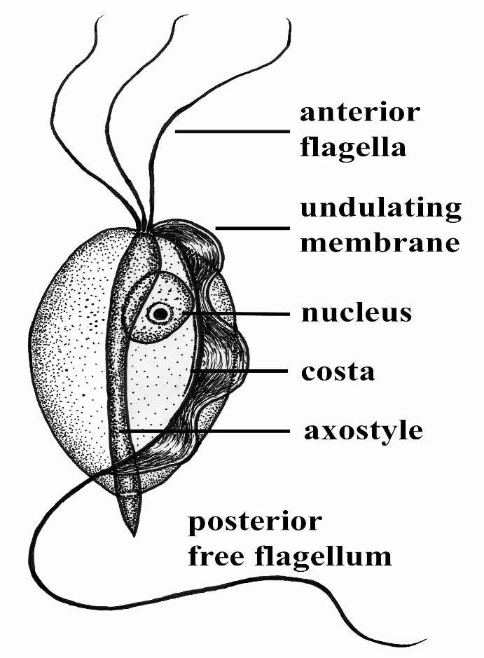
The T. foetus , like other trichomonads, has only trophozoite stage, however, it has a pseudocyst stage (Lipman NS, et. al., 1999) (Pereira-Neves, Ribeiro et al. 2003, Benchimol 2004, Mariante, Lopes et al. 2004, Yao and Köster 2015). Therefore, cats are presumed to be infected via direct contact due to the absence of the cyst stage (Zajac AM, Conboy GA, 2006). The parasite is pear- or spindle-shaped, having three anterior flagella and one posterior flagellum. With its size of approximately 10-25 μm in length and 3-15 μm in width, the undulating membrane extends along the whole length of the body which emerges as the posterior flagellum. This trophozoite reproduces asexually by longitudinal binary fission (Yao and Köster 2015).
(https://www.k-state.edu/parasitology/625tutorials/Protozoa10.html)
Fig. 01. Morphologic structure of the Tritrichomonas foetus
Infected cats were reported to experience chronic large bowel diarrhea as the parasite was found in the feline intestine (Foster, Gookin et al. 2004, Payne and Artzer 2009). Reports also showed the presence of the organism in the feline uterus (Dahlgren, Gjerde et al. 2007). Further, the isolates of T. foetus from cattle are known to be infectious for the cats, while the isolates of the same species from cats are infectious to cattle (Stockdale, Dillon et al. 2008, Walden, Rodning et al. 2008, Payne and Artzer 2009).
Clinical Signs/ Pathogenesis
Despite suggestions of strong association of feline T. foetus and chronic diarrhea (Gookin, Stebbins et al. 2004, Mardell and Sparkes 2006, Gunn-Moore, McCann et al. 2007, Burgener, Frey et al. 2009, Holliday, Deni et al. 2009, Pham 2009, Stockdale, Givens et al. 2009, Kuehner, Marks et al. 2011), whether the parasite alone is sufficient to cause clinical signs or the foetus-associated diarrhea, being a primarily multifactorial disease involves concurrent infection with other enteropathogens, host and environmental factors (Gookin, JL, et. al., 1999) (Gookin, Levy et al. 2001, Bissett, Gowan et al. 2008, Stockdale, Givens et al. 2009, Kuehner, Marks et al. 2011).
It is possible that the trophozoites are transmitted by a fecal- oral route from an infected to uninfected cat (Yao and Köster 2015). Symptoms may appear early as 2 to 7 days after orogastric inoculation (Gookin, Levy et al. 2001, Yao and Köster 2015). Whereas, infected cats showed anorexia, depression, vomiting and weight loss while experimental infections also reported vomiting and fever (Mardell and Sparkes 2006, Xenoulis, Lopinski et al. 2013, Yao and Köster 2015)( Stockdale, H., et al., 2007). Chronic large bowel diarrhea, associated with blood, mucus, flatulence, tenesmus, and anal irritation were also reported (Foster, Gookin et al. 2004, Gookin, Stebbins et al. 2004, Payne and Artzer 2009, Stockdale, Givens et al. 2009). With the characteristic of the trichomonads as commensal organisms, some hosts show no clinical signs and are asymptomatic (Payne and Artzer 2009).
In some studies, the parasite, with its surface located antigen, was detected on epithelial surface and within the superficial detritus of the cecal and colonic mucosa (Gookin, Levy et al. 2001, Yao and Köster 2015), while naturally infected cats showed parasite in close proximity to the mucosal surface and less frequently in the lumen of colonic crypts (Yaeger and Gookin 2005, Yao and Köster 2015). Conclusively, T. foetus trophozoites can be detected in epithelial surface and crypts of cecum and colon (Yao and Köster 2015). Mechanisms were then described to include possibilities of alterations in the normal intestinal flora, adherence to the epithelium, and elaboration of cytokines and enzymes (Payne and Artzer 2009).
Diagnosis
For cats <6months old with recent clinical signs of chronic large bowel diarrhea, infection with T. foetus infection is suspected (Yao and Köster 2015).
Diagnosis of the T. foetus infection may be conducted via direct observation of the flagellates in fresh or cultured feces (Payne and Artzer 2009) or on a saline diluted direct fecal smear (Yao and Köster 2015). The trophozoites of T. foetus are difficult to distinguish from Giardia spp and other nonpathogenic intestinal trichomonads. However, although the size of T. foetus and Giardia spp are almost the same, they move differently. The movement of Giardia spp. resembles the fall of a leaf while trichomonads move erratically (Yao and Köster 2015)( Gookin JL, Levy MG, 2008).
Feline feces can be cultivated and be tested in commercially available InPouch™ TF medium (Payne and Artzer 2009, Yao and Köster 2015) or DNA extraction and amplification of T. foetus rDNA by the use of PCR from feces samples can be conducted (Gookin, Stebbins et al. 2004, Manning 2010, Yao and Köster 2015). Moreover, other causes of diarrhea, like bacterial, viral, other parasites, and nutritional problems should be ruled out before a diagnosis of tritrichomoniasis can be made (Payne and Artzer 2009).
Treatment and Disease Managements
The T. foetus infection in cats has no approved treatment. While treating infected animals is difficult, success is also limited (Payne and Artzer 2009). Literatures have used therapeutics including paromomycin, fenbendazole, furazolidone, nitazoxanide, metronidazole, tinidazole and ronidazole (Gookin, Breitschwerdt et al. 1999, Gookin, Levy et al. 2001, Yao and Köster 2015).
The ronidazole is not registered for human or veterinary use (Yao and Köster 2015) however, it showed effectiveness in experimentally infected cats (Gookin, Copple et al. 2006) (Gookin JL, Dybas D, 2008). Due to possible neurologic side effects, caution is observed in using this drug. Moreover, relieving diarrheal symptoms such as dietary changes have also failed to help resolve symptoms (Payne and Artzer 2009).
Therefore, sanitation of the environment and the animals in a cattery is critical. This parasite do not survive outside the host for a long time, but cats are fastidious and will definitely re-ingest these parasites readily (Payne and Artzer 2009).
References:
Gookin JL, Dybas D. An owners guide to diagnosis and treatment of cats infected with Tritrichomonas foetus. Available at: http://www.cvm.ncsu.edu/docs/ documents/ownersguide_tfoetus_revised042808.pdf. Accessed April 28, 2008.
Gookin JL, Levy MG. Discrimination of Tritrichomonas foetus and Giardia by light microscopy. www. 2008. Ref Type: Electronic Citation.
Stockdale H, Rodning S, Givens M, Carpenter D, Lenz S, Spencer J, Dykstra C, Lindsay D, Blagburn B (2007) Experimental infection of cattle with a feline isolate of Tritrichomonas foetus. J Parasitol 93:1429–1434
Zajac AM, Conboy GA. Fecal examination for the diagnosis of parasitism. Veterinary clinical parasitology. 7th edition. Ames (IA): Blackwell; 2006. p. 3–148
Levy MG, Gookin JL, Poore MF, Dykstra M, Litaker RW. Tritrichomonas foetus and not Pentatrichomonas hominis is the causative agent of the recently described feline intestinal trichomonosis. J Vet Intern Med 2001; 15: 315.
Gookin JL, Breitschwerdt EB, Levy MG, Gager RB, Benrud JG. Diarrhea associated with trichomonosis in cats. J Am Vet Med Assoc 1999; 215: 1450e4.
Lipman NS, Lampen N, Nguyen HT (1999) Identification of pseudocysts of Tritrichomonas muris in Armenian hamsters and their transmission to mice. Lab Anim Sci 49:313–315
Gunn-Moore D, Tennant B. Tritrichomonas foetus diarrhoea in cats. Vet Rec 2007; 160: 850
Bell, E. T., R. A. Gowan, A. E. Lingard, R. J. McCoy, J. Slapeta and R. Malik (2010). “Naturally occurring Tritrichomonas foetus infections in Australian cats: 38 cases.” J Feline Med Surg 12(12): 889-898.
Bell, E. T., R. A. Gowan, A. E. Lingard, R. J. McCoy, J. Šlapeta and R. Malik (2010). “Naturally occurring Tritrichomonas foetus infections in Australian cats: 38 cases.” Journal of Feline Medicine and Surgery 12(12): 889-898.
Benchimol, M. (2004). “Trichomonads under Microscopy.” Microsc Microanal 10(5): 528-550.
Bissett, S. A., R. A. Gowan, C. R. O’Brien, M. R. Stone and J. L. Gookin (2008). “Feline diarrhoea associated with Tritrichomonas cf. foetus and Giardia co-infection in an Australian cattery.” Aust Vet J 86(11): 440-443.
Burgener, I., C. Frey, P. Kook and B. Gottstein (2009). “[Tritrichomonas fetus: a new intestinal parasite in Swiss cats].” Schweiz Arch Tierheilkd 151(8): 383-389.
Dahlgren, S. S., B. Gjerde and H. Y. Pettersen (2007). “First record of natural Tritrichomonas foetus infection of the feline uterus.” J Small Anim Pract 48(11): 654-657.
Foster, D. M., J. L. Gookin, M. F. Poore, M. E. Stebbins and M. G. Levy (2004). “Outcome of cats with diarrhea and Tritrichomonas foetus infection.” J Am Vet Med Assoc 225(6): 888-892.
Frey, C. F., M. Schild, A. Hemphill, P. Stünzi, N. Müller, B. Gottstein and I. A. Burgener (2009). “Intestinal Tritrichomonas foetus infection in cats in Switzerland detected by in vitro cultivation and PCR.” Parasitol Res 104(4): 783-788.
Gookin, J. L., E. B. Breitschwerdt, M. G. Levy, R. B. Gager and J. G. Benrud (1999). “Diarrhea associated with trichomonosis in cats.” J Am Vet Med Assoc 215(10): 1450-1454.
Gookin, J. L., C. N. Copple, M. G. Papich, M. F. Poore, S. H. Stauffer, A. J. Birkenheuer, D. C. Twedt and M. G. Levy (2006). “Efficacy of ronidazole for treatment of feline Tritrichomonas foetus infection.” J Vet Intern Med 20(3): 536-543.
Gookin, J. L., M. G. Levy, J. M. Law, M. G. Papich, M. F. Poore and E. B. Breitschwerdt (2001). “Experimental infection of cats with Tritrichomonas foetus.” Am J Vet Res 62(11): 1690-1697.
Gookin, J. L., S. H. Stauffer, D. Dybas and D. H. Cannon (2010). “Documentation of In Vivo and In Vitro Aerobic Resistance of Feline Tritrichomonas foetus Isolates to Ronidazole.” Journal of Veterinary Internal Medicine 24(4): 1003-1007.
Gookin, J. L., M. E. Stebbins, E. Hunt, K. Burlone, M. Fulton, R. Hochel, M. Talaat, M. Poore and M. G. Levy (2004). “Prevalence of and risk factors for feline Tritrichomonas foetus and giardia infection.” Journal of clinical microbiology 42(6): 2707-2710.
Gunn-Moore, D. A., T. M. McCann, N. Reed, K. E. Simpson and B. Tennant (2007). “Prevalence of Tritrichomonas foetus infection in cats with diarrhoea in the UK.” J Feline Med Surg 9(3): 214-218.
Holliday, M., D. Deni and D. A. Gunn-Moore (2009). “Tritrichomonas foetus infection in cats with diarrhoea in a rescue colony in Italy.” J Feline Med Surg 11(2): 131-134.
Kuehner, K. A., S. L. Marks, P. H. Kass, C. Sauter-Louis, R. A. Grahn, D. Barutzki and K. Hartmann (2011). “Tritrichomonas foetus infection in purebred cats in Germany: Prevalence of clinical signs and the role of co-infection with other enteroparasites.” Journal of Feline Medicine and Surgery 13(4): 251-258.
Levy, M. G., J. L. Gookin, M. Poore, A. J. Birkenheuer, M. J. Dykstra and R. W. Litaker (2003). “Tritrichomonas foetus and not Pentatrichomonas hominis is the etiologic agent of feline trichomonal diarrhea.” J Parasitol 89(1): 99-104.
Manning, K. (2010). “Update on the diagnosis and management of Tritrichomonas foetus infections in cats.” Top Companion Anim Med 25(3): 145-148.
Mardell, E. J. and A. H. Sparkes (2006). “Chronic diarrhoea associated with Tritrichomonas foetus infection in a British cat.” Vet Rec 158(22): 765-766.
Mariante, R. M., L. C. Lopes and M. Benchimol (2004). “Tritrichomonas foetus pseudocysts adhere to vaginal epithelial cells in a contact-dependent manner.” Parasitol Res 92(4): 303-312.
Payne, P. A. and M. Artzer (2009). “The biology and control of Giardia spp and Tritrichomonas foetus.” Vet Clin North Am Small Anim Pract 39(6): 993-1007, v.
Pereira-Neves, A., K. C. Ribeiro and M. Benchimol (2003). “Pseudocysts in trichomonads–new insights.” Protist 154(3-4): 313-329.
Pham, D. (2009). “Chronic intermittent diarrhea in a 14-month-old Abyssinian cat.” The Canadian veterinary journal = La revue veterinaire canadienne 50(1): 85-87.
Stockdale, H. D., A. R. Dillon, J. C. Newton, R. C. Bird, R. H. Bondurant, P. Deinnocentes, S. Barney, J. Bulter, T. Land, J. A. Spencer, D. S. Lindsay and B. L. Blagburn (2008). “Experimental infection of cats (Felis catus) with Tritrichomonas foetus isolated from cattle.” Vet Parasitol 154(1-2): 156-161.
Stockdale, H. D., M. D. Givens, C. C. Dykstra and B. L. Blagburn (2009). “Tritrichomonas foetus infections in surveyed pet cats.” Veterinary parasitology 160(1-2): 13-17.
Walden, H., S. Rodning, M. Givens, D. Carpenter, S. Lenz, J. Spencer, C. Dykstra, D. Lindsay and B. Blagburn (2008). “Experimental infection of cattle with a feline isolate of Tritrichomonas foetus.” The Journal of parasitology 93: 1429-1434.
Xenoulis, P. G., D. J. Lopinski, S. A. Read, J. S. Suchodolski and J. M. Steiner (2013). “Intestinal Tritrichomonas foetus infection in cats: a retrospective study of 104 cases.” J Feline Med Surg 15(12): 1098-1103.
Yaeger, M. J. and J. L. Gookin (2005). “Histologic features associated with tritrichomonas foetus-induced colitis in domestic cats.” Vet Pathol 42(6): 797-804.
Yao, C. and LS Köster (2015). “Tritrichomonas foetus infection, a cause of chronic diarrhea in the domestic cat.” Veterinary Research 46 (1): 35.