
Feline Herpesvirus 1 (FHV-1) Infection
Bioguard Corporation Feline herpesvirus-1 (FHV-1) is a common viral infection in cats that primarily affects the upper respiratory system. It is a major cause of feline viral rhinotracheitis (FVR), which presents symptoms like sneezing, nasal discharge, and conjunctivitis (eye inflammation). FHV-1 is highly contagious among cats and is spread through direct contact with infected saliva, eye or nasal secretions, or contaminated environments. Once infected, many cats can become lifelong carriers of the virus, with symptoms reoccurring during times of stress or illness. Transmission A cat can become infected with this virus through direct exposure to viral particles. The virus is transmitted through saliva, as well as eye and nasal discharges from an infected cat. Infection occurs when a susceptible cat comes into contact with an infected cat or with contaminated objects, such as clothing, food and water bowls, or furniture that carry the viral particles. Though the virus is fragile in the environment and doesn’t survive for long outside the host, it remains a significant concern because many cats become lifelong carriers after infection. Even when no symptoms are present, stress or illness can trigger a flare-up, allowing the carrier to shed the virus and infect others. Incubation Period of FHV-1 Infection After a cat is infected with FVR, symptoms typically appear within two to five days, which is the incubation period of the disease. During this time, the cat can already infect other cats. Once symptoms develop, the active infection usually lasts about 10 to 20 days. Clinical Signs Upper respiratory signs of FVR include sneezing, nasal discharge, fever, loss of appetite, and coughing. Eye-related symptoms may encompass discharge, conjunctivitis or chemosis, changes in color, and corneal ulcers. In severe cases, the skin around the face may show signs such as redness, swelling, crusting, and hair loss. Other non-specific symptoms can include fever, lethargy (tiredness), anorexia (poor appetite), and enlarged lymph nodes. Diagnosis In most cases, a specific diagnosis of FHV infection is not necessary. A presumptive diagnosis of FVR is based primarily on a cat’s medical history and clinical signs combined with the findings on physical examination, particularly if the cat has evidence of a corneal infection. Corneal staining with fluorescein dye is often performed to look for any ulcers that may have developed. However, if a specific diagnosis is needed, ocular or oral swabs can be sent to a veterinary lab. There, the virus can be cultured or, more commonly detected by PCR. Additionally, evidence of the virus can be found in biopsies, which can help diagnose FHV-associated dermatitis. Prevention Preventing FVR involves several key strategies: Vaccination: Core vaccines for cats include the FVR vaccine, which significantly reduces the severity and duration of the illness, even if it doesn’t completely prevent infection. Hygiene and Sanitation: Good hygiene practices, such as washing your hands thoroughly before and after handling other cats, can help prevent the spread of FHV-1. Keeping your cat’s living environment clean is also important. Minimizing Stress: Stress can trigger the reactivation of the virus in carrier cats. Providing a stable, stress-free environment can help reduce the likelihood of outbreaks. Isolation: If the cat is infected, keeping her isolated from other cats can prevent the spread of the virus. This is especially important in multi-cat households, boarding facilities, and shelters. References Bergmann M, Speck S, Rieger A, et al. Antibody response to feline herpesvirus-1 vaccination in healthy adult cats. J Feline Med Surg. 2020 Apr;22(4):329-338. Cottingham E, Johnstone T, Hartley CA, et al. Update on feline alphaherpesvirus-1 seroprevalence in Victorian feral and owned cats. Aust Vet J. 2022 May;100(5):187-189. Thomasy SM, Maggs DJ. A review of antiviral drugs and other compounds with activity against feline herpesvirus type 1. Vet Ophthalmol. 2016 Jul;19 Suppl 1(Suppl 1):119-30.
Feline Herpesvirus Infection- Diagnosis
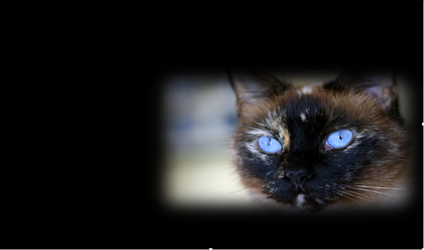
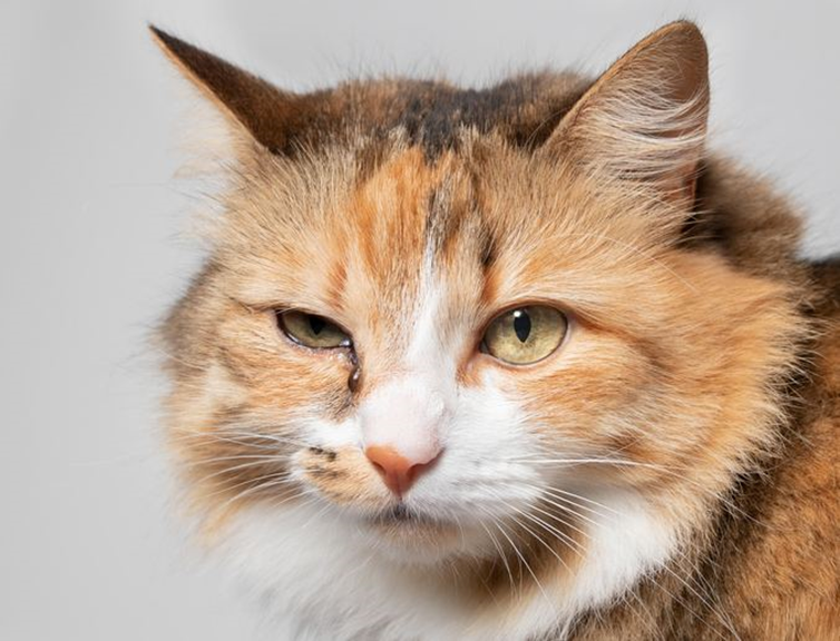
Trinh Mai Nguyen Tang Feline herpesvirus-1 (FHV-1) is a feline respiratory infection virus also known as feline viral rhinotracheitis (FVR) [1]. The Herpes virus was first isolated by scientists Crandell and Maurer in 1958 in cats with respiratory infections [2]. This virus has a prominent genome with large double stranded DNA, belonging to the family Herpesviridae [3]. This virus is characterized by cat-to-cat transmission with an exposure rate of up to 97% [4]. Herpes virus can be inactivated at 37oC around 3 hours or at 56oC in 5 mins. Meanwhile, the virus can remains infective in the enviroment approximately 5 months and a month at 25oC [5]. Once a cat is infected with the herpes virus, it is incredibly difficult to completely treat it since the virus can enter a dormant state and continue to survive in the cat for the remainder of its life [6]. Cats are not infectious during this latent period, but if they are sick or going through a stressful period, the virus may be reactivate. If this occurs, the cat will once more get the infection and may represent symptoms [7]. Herpes viruses can be latent in the ganglion, attach to sensory nerves and reach nerve cells, persist in the nucleus of infected nerve cells and do not replicate, leading to the process of detecting this virus becomes difficult [7-8]. As reported by Ngoc.N.T and her colleagues, herpes virus can infect cats of any age, however, kittens are more susceptible [9]. Specifically, the prevalence of virus infection in cats younger than 6 months old, 6-12 months old and over 12 months old were 52.17%, 33.33% and 19.05%, respectively [9]. Although previous reports have demonstrate that gender has no effect on the incidence of herpes virus infection [7-8], but Henzel et al. (2002) found that isolates from female cats are substantially taller than isolates from male cats [10]. Clinical Symptoms Herpes virus enters the cat’s body by contact with infected tears, nose, saliva, or items, and then multiplies rapidly in the epithelium of the nose, nasopharynx, and conjunctival mucosa leading to primary infection [11]. In cats infected with FHV-1, signs of sadness, moodiness, lethargy, sneezing, fever, and discharge from the eyes and nose have been noted (figure 1-A), a process that is frequently extended 2-4 days or longer, depending on the immunological system of the cat [11]. Secondary infection occurred after the fourth day of incubation, with symptoms of infection in the throat, bronchi, and bronchioles, and the nasal and conjunctival epithelium necrosing [12]. Conjunctivitis is a common herpes virus symptom, indicated by congestive and exudative symptoms that develop over many days to purulent discharge (figure 1-B) [13]. Gaskell and Dawson (1988) found lung infection or bronchitis in cats, with kittens dying from pneumonia at a greater incidence than adult cats [11,14]. Some other atypical symptoms such as mouth and skin ulcers, dermatitis or neurological signs are rarely seen [7]. Furthermore, the mean white blood cell (WBC) count of cats infected with FHV-1 (17.77 ± 0.70 x10³/μl) was slightly increased compared with that of normal cats (4.6-12.8 x 10³/μl) [9], in which neutrophils, eosinophils and monocytes all showed signs of slight increase compared. Secondary infections of the eyes, upper respiratory tract, and necrotic ulcers of the mouth can all cause high white blood cell counts [11]. Table 1 displays the white blood cell count. Table 1. Hematological results of cats was infected with FHV-1 [9]. Targets Unit Reference ± SE Red blood cells ´ 106/μl 7-10,7 10,20 ± 0,64 Hemoglobin content g/dl 11,3-15,5 13,47 ± 0,54 RBC mass % 33-45 38,78 ± 1,41 Average volume of red blood cells fl 41-49 45,16 ± 0,75 Average amount of hemoglobin in red blood cells pg 14-17 15,57 ± 0,24 Platelet count ´ 103/μl 180-680 362,50 ± 30,82 WBC count ´ 103/μl 4,6-12,8 17,77 ± 0,70 Lymphocytes ´ 103/μl 1,05-6,00 4,50 ± 0,36 Mono leukocytes ´ 103/μl 0,05-0,68 0,96 ± 0,13 Neutrophils polymorphonuclear leukocytes ´ 103/μl 2,32-10,01 11,47 ± 0,45 Eosinophils ´ 103/μl 0,1-0,6 0,78 ± 0,08 Basophils ´ 103/μl 0-0,14 0,07 ± 0,01 Laboratory Diagnosis In the laboratory, there are many different methods used to determine FHV. Common approaches include PCR, virus isolation in cell culture, and indirect fluorescent antibody staining of tissue samples for viral antibody detection [8,16]. PCR FHV is one of the most common causes of upper respiratory tract illness in cats. Infected cats would show upper respiratory signs. Co-infection of FHV with other pathogens makes the clinical signs more severe, particularly feline calicivirus, Chlamydophila felis, Bordetella pneumoniaseptica, Mycoplasma species, Staphylococcus spp., or Escherichia coli [6]. When a cat is suspected of having a viral infection, a swab can be used to collect nasal, ocular, oropharyngeal secretions, corneal debris, aqueous humor, corneal samples, blood, or biopsies. By amplifying viral DNA, tPCR can detect genetic material of FHV in specimen. However, if the cat is not in the infectious phase, no virus particles will be shed, rendering the PCR test ineffective [19-20]. ELISA method – detecting IgG antibodies The enzyme-linked immunosorbent test (ELISA) is used to determine IgG antibodies against FHV or FHV-1 utilizing serum, aqueous humor, and cerebrospinal fluid samples [15]. This approach, however, cannot discriminate between diseased and vaccinated cats. Due to the extended latent period of FHV, the cat’s body will generate antibodies to combat it [8]. These neutralizing antibodies manifest 20-30 days after the first infection. As a result, the presence of antibodies in the serum signals a prior infection but does not always correspond with clinical signs [8]. Immunofluorescent antibody assay Another approach for detecting FHV is immunofluorescence antibody (IFA) testing on corneal or conjunctival smears or biopsiengs. Through an antigen-antibody response, this assay may identify viral proteins produced in cells. However, this approach is thought to be less sensitive than viral isolation or PCR [16]. Virus isolation This is a traditional method that can detect viruses through isolation of conjunctival debris, nose, oropharynx, or postmortem lung samples from infected cats [8]. This traditional method can detect