
Maigan Espinili Maruquin
The lipases are present from a variety of cells, including pancreatic, hepatic, and gastric cells, with similar function, ie , hydrolysis of triglycerides (Xenoulis and Steiner 2012)( Steiner JM, 2000)(Dröes and Tappin 2017). From the pancreatic origin, the canine pancreatic lipase increases in the event of pancreatic inflammation (Steiner and Williams 2003, Haworth, Hosgood et al. 2014).
The canine pancreatic lipase immunoreactivity (cPLI) assay was first a radioimmunoassay, and subsequently an enzyme immunoassay, eventually developed into a commercially available specific canine pancreatic lipase assay (Steiner, Teague et al. 2003, Steiner and Williams 2003, Haworth, Hosgood et al . 2014). The serum cPLI is now used as an important specific, and sensitive marker for the exocrine pancreas (Steiner, Newman et al. 2008, Neilson-Carley, Robertson et al. 2011, Trivedi, Marks et al. 2011, Xenoulis and Steiner 2012, Mawby, Whittemore et al. 2014, Dröes and Tappin 2017).
Canine Pancreatitis
Disorders originating from the liver and pancreas are considered important causes of morbidity and mortality in both dogs and cats, which presents different sets of challenges in diagnosis (Lidbury and Suchodolski 2016). Dogs have several exocrine pancreas diseases including exocrine pancreatic insufficiency (EPI), pancreatic carcinoma, and pancreatitis (Lidbury and Suchodolski 2016). Among them, pancreatitis is the most common disorder and are mostly considered idiopathic (Xenoulis and Steiner 2012).
Pancreatitis is the inflammation of the exocrine pancreas, wherein there is an infiltration with inflammatory cells. The term is usually expanded to include other diseases of the exocrine pancreas by necrosis or necrotising pancreatitis, or irreversible structural changes such as fibrosis (chronic pancreatitis) (Xenoulis and Steiner 2012). The pancreatitis is in acute stage when there is neutrophilic inflammation while chronic is characterized by the acinar atrophy and fibrosis (Newman, Steiner et al. 2006, Watson, Roulois et al. 2007, Mansfield, Anderson et al. 2012 , Watson 2015, Dröes and Tappin 2017).
 https://i2.wp.com/thewholedog.com/wp-content/uploads/2014/04/pancreatiitis.jpg?ssl=1
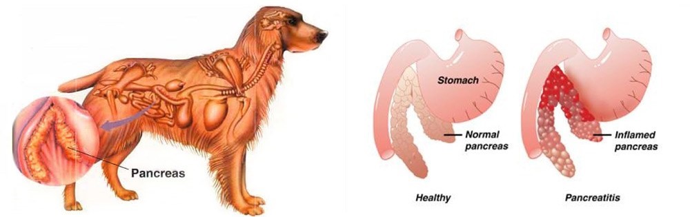
Fig. 01. An illustration of the difference of healthy pancreas vs. inflamed pancreas in dogs
Despite being a common disorder in canines, the antemortem diagnosis of pancreatitis is clinically challenging (Trivedi, Marks et al. 2011). Clinical signs are non-specific and varies greatly. Subclinical diseases are evident in most cases while others may display mild and non -specific clinical signs (Xenoulis 2015) which may include anorexia, vomiting, lethargy, diarrhea, melena, weight loss, hematemesis, and hematochezia (Hess, Saunders et al. 1998, Trivedi, Marks et al. 2011). Dogs with acute pancreatitis are at risk of developing obesity, diabetes mellitus, hyperadrenocortcisim, hypothyroidism, prior gastrointestinal disease, and epilepsy (Cook, Breitschwerdt et al. 1993, Trivedi, Marks et al. 2011). On the other hand, cardiovascular shock,disseminated intravascular coagulation (DIC) or multi-organ failure and death within hours of the development of clinical signs are presented in severe cases (Xenoulis 2015, Dröes and Tappin 2017).
As diagnosis of pancreatitis remains a challenge, histopathology is considered the gold standard for diagnosis of pancreatitis. However, with procedural risks and difficulty to determine the right area for biopsy, obtaining the samples is hard (Newman, Steiner et al. 2004, Dröes and Tappin 2017). Therefore, considering the improving sensitivity and specificity of laboratory tests, using a combination of different methods can be best used (Dröes and Tappin 2017).
On the other hand, there are treatments for acute pancreatitis infected dogs. Treatments consider Intravenous Fluid Therapy, knowing that pancreatitis disturbs pancreatic microcirculation (Bassi, Kollias et al. 1994, Mansfield 2012). The administration of plasma is claimed to correct hypoalbuminemia, replacement of circulating α-macroglobulins, replacement of coagulation factors, and amelioration of systemic inflammation, however, no controlled studies on plasma transfusion in dogs with naturally occurring acute pancreatitis has proven its benefit (Mansfield 2012).
Due to vomiting of infected dogs, anti-emetics are used to manage acute pancreatitis (Mansfield 2012). On the other hand, corticosteroids enhance apoptosis, and increase the production of pancreatitis-associated protein, giving protective effect against pancreatic inflammation (Zhang, Kandil et al. 2004, Mansfield 2012). Moreover, diet should also be managed including the treatment of the complications of the disease (Mansfield 2012)
Canine Pancreatic Lipase Immunoreactivity Assay
Due to the limitations of the traditional golden standard, histopathology, diagnosis has relied on clinical criteria as an alternative gold standard (Graca, Messick et al. 2005, McCord, Morley et al. 2012, Cridge, MacLeod et al. 2018, Gori, Lippi et al. 2019, Nielsen, Holm et al. 2019, Okanishi, Nagata et al. 2019, Cridge, Mackin et al. 2020) and this includes measurement of cPLI (Cridge, Mackin et al. 2020). It has been proven over the decade that the PLI assay development, analytical validation, and evaluation is very useful for the diagnosis of pancreatitis in both dogs and cats (Xenoulis and Steiner 2012)( Xenoulis PG, Steiner JM, 2013).
Pancreatic lipase is expressed exclusively by pancreatic acinar cells and thus plays an important role in assessing the exocrine pancreas (Steiner, Berridge et al. 2002, Steiner, Rutz et al. 2006, Neilson-Carley, Robertson et al. 2011, Xenoulis and Steiner 2012). The serum cPLI has been reported to detect specifically localized lipase in pancreatic acinar cells (Steiner, Berridge et al. 2002, Dröes and Tappin 2017). This makes increase in serum PLI concentrations unlikely coming from the lipase of other tissues (Lidbury and Suchodolski 2016). Considering the localization of the pancreatic lipase and the capacity of immunoassays to detect the unique protein structure of pancreatic lipase are known to be the advantages of measuring the serum cPLI during the diagnosis of exocrine pancreas diseases, resulting to high analytic specificity (Steiner,Rutz et al. 2006, Xenoulis and Steiner 2012) (Steiner JM, 2000; Hoffmann WE, 2008) (Steiner, Berridge et al. 2002, Neilson-Carley, Robertson et al. 2011).
Nowadays, diagnosis of canine pancreatitis has mostly considered serum cPLI assays as the most specific serum biomarkers (Neilson-Carley, Robertson et al. 2011, Trivedi, Marks et al. 2011, Mawby, Whittemore et al. 2014). However, the serum cPLI concentration doesn’t define the severity of pancreatitis (Steiner, Newman et al. 2008, Trivedi, Marks et al. 2011, Xenoulis and Steiner 2012).
References:
Hoffmann WE. Diagnostic enzymology of domestic animals. In: Kaneko JJ, Harvey JW, Bruss ML, eds. Clinical Biochemistry of Domestic Animals. 6th ed. Burlington, MA: Academic Press; 2008:351–378
Steiner JM. Canine digestive lipases. PhD Thesis. Texas A&M University; 2000:1–251.
Xenoulis PG, Steiner JM. Diagnostic evaluation of the pancreas. In: Washabau RJ, Day MJ, eds. Canine and Feline Gastroenterology. St. Louis, MO: Elsevier; 2013: 803–812.
Bassi, D., N. Kollias, C. Fernandez-del Castillo, T. Foitzik, AL Warshaw and DW Rattner (1994). “Impairment of pancreatic microcirculation correlates with the severity of acute experimental pancreatitis.” J Am Coll Surg 179 ( 3): 257-263.
Cook, AK, EB Breitschwerdt, JF Levine, SE Bunch and LO Linn (1993). “Risk factors associated with acute pancreatitis in dogs: 101 cases (1985-1990).” J Am Vet Med Assoc 203 (5): 673- 679.
Cridge, H., AJ Mackin, JA Lidbury, JS Suchodolski and JM Steiner (2020). “Comparative repeatability of pancreatic lipase assays in the commercial and in-house laboratory environments.” J Vet Intern Med 34 (3): 1150-1156 .
Cridge, H., AG MacLeod, GE Pachtinger, AJ Mackin, AM Sullivant, JM Thomason, TM Archer, KV Lunsford, K. Rosenthal and RW Wills (2018). “Evaluation of SNAP cPL, Spec cPL, VetScan cPL Rapid Test, and Precision PSL Assays for the Diagnosis of Clinical Pancreatitis in Dogs.” J Vet Intern Med 32 (2): 658-664.
Dröes, F. and S. Tappin (2017). “Canine pancreatitis — a challenging disease. Part 1.” Companion Animal 22 (4): 224-232.
Gori, E., I. Lippi, G. Guidi, F. Perondi, A. Pierini and V. Marchetti (2019). “Acute pancreatitis and acute kidney injury in dogs.” Vet J 245 : 77-81.
Graca, R., J. Messick, S. McCullough, A. Barger and W. Hoffmann (2005). “Validation and diagnostic efficacy of a lipase assay using the substrate 1,2-o-dilauryl-rac-glycero glutaric acid- (6′ methyl resorufin)-ester for the diagnosis of acute pancreatitis in dogs.” Vet Clin Pathol 34 (1): 39-43.
Haworth, MD, G. Hosgood, KL Swindells and CS Mansfield (2014). “Diagnostic accuracy of the SNAP and Spec canine pancreatic lipase tests for pancreatitis in dogs presenting with clinical signs of acute abdominal disease.” Journal of Veterinary Emergency and Critical Care 24 (2): 135-143.
Hess, RS, HM Saunders, TJ Van Winkle, FS Shofer and RJ Washabau (1998). “Clinical, clinicopathologic, radiographic, and ultrasonographic abnormalities in dogs with fatal acute pancreatitis: 70 cases (1986-1995).” J Am Vet Med Assoc 213 (5): 665-670.
Lidbury, JA and JS Suchodolski (2016). “New advances in the diagnosis of canine and feline liver and pancreatic disease.” The Veterinary Journal 215 : 87-95.
Mansfield, C. (2012). “Acute Pancreatitis in Dogs: Advances in Understanding, Diagnostics, and Treatment.” Topics in Companion Animal Medicine 27 (3): 123-132.
Mansfield, CS, GA Anderson and AJ O’Hara (2012). “Association between canine pancreatic-specific lipase and histologic exocrine pancreatic inflammation in dogs: assessing specificity.” J Vet Diagn Invest 24 (2): 312-318.
Mawby, DI, JC Whittemore and KA Fecteau (2014). “Canine Pancreatic-Specific Lipase Concentrations in Clinically Healthy Dogs and Dogs with Naturally Occurring Hyperadrenocorticism.” Journal of Veterinary Internal Medicine 28 (4): 1244-1250.
McCord, K., PS Morley, J. Armstrong, K. Simpson, M. Rishniw, MA Forman, D. Biller, N. Parnell, K. Arnell, S. Hill, S. Avgeris, H. Gittelman, M. Moore , M. Hitt, G. Oswald, S. Marks, D. Burney and D. Twedt (2012). “A multi-institutional study evaluating the diagnostic utility of the spec cPL™ and SNAP® cPL™ in clinical acute pancreatitis in 84 dogs.” J Vet Intern Med 26 (4): 888-896.
Neilson-Carley, SC, JE Robertson, SJ Newman, D. Kutchmarick, R. Relford, K. Woosley and JM Steiner (2011). “Specificity of a canine pancreas-specific lipase assay for diagnosing pancreatitis in dogs without clinical or histologic evidence of the disease.” Am J Vet Res 72 (3): 302-307.
Newman, S., J. Steiner, K. Woosley, L. Barton, C. Ruaux and D. Williams (2004). “Localization of pancreatic inflammation and necrosis in dogs.” J Vet Intern Med 18 (4): 488- 493.
Newman, SJ, JM Steiner, K. Woosley, DA Williams and L. Barton (2006). “Histologic assessment and grading of the exocrine pancreas in the dog.” J Vet Diagn Invest 18 (1): 115-118.
Nielsen, L., J. Holm, E. Rozanski, D. Meola, LL Price and A. de Laforcade (2019). “Multicenter investigation of hemostatic dysfunction in 15 dogs with acute pancreatitis.” J Vet Emerg Crit Care (San Antonio ) 29 (3): 264-268.
Okanishi, H., T. Nagata, S. Nakane and T. Watari (2019). “Comparison of initial treatment with and without corticosteroids for suspected acute pancreatitis in dogs.” J Small Anim Pract 60 (5): 298-304.
Steiner, JM, BR Berridge, J. Wojcieszyn and DA Williams (2002). “Cellular immunolocalization of gastric and pancreatic lipase in various tissues obtained from dogs.” American Journal of Veterinary Research 63 (5): 722-727.
Steiner, JM, BR Berridge, J. Wojcieszyn and DA Williams (2002). “Cellular immunolocalization of gastric and pancreatic lipase in various tissues obtained from dogs.” Am J Vet Res 63 (5): 722-727.
Steiner, JM, S. Newman, P. Xenoulis, K. Woosley, J. Suchodolski, D. Williams and L. Barton (2008). “Sensitivity of serum markers for pancreatitis in dogs with macroscopic evidence of pancreatitis.” Vet Ther 9 (4): 263-273.
Steiner, JM, GM Rutz and DA Williams (2006). “Serum lipase activities and pancreatic lipase immunoreactivity concentrations in dogs with exocrine pancreatic insufficiency.” Am J Vet Res 67 (1): 84-87.
Steiner, JM, SR Teague and DA Williams (2003). “Development and analytic validation of an enzyme-linked immunosorbent assay for the measurement of canine pancreatic lipase immunoreactivity in serum.” Canadian journal of veterinary research = Revue canadienne de recherche veterinaire 67 ( 3): 175-182.
Steiner, JM and DA Williams (2003). “Development and validation of a radioimmunoassay for the measurement of canine pancreatic lipase immunoreactivity in serum of dogs.” Am J Vet Res 64 (10): 1237-1241.
Trivedi, S., SL Marks, PH Kass, JA Luff, SM Keller, EG Johnson and B. Murphy (2011). “Sensitivity and Specificity of Canine Pancreas-Specific Lipase (cPL) and Other Markers for Pancreatitis in 70 Dogs with and without Histopathologic Evidence of Pancreatitis.” Journal of Veterinary Internal Medicine 25 (6): 1241-1247.
Watson, P. (2015). “Pancreatitis in dogs and cats: definitions and pathophysiology.” J Small Anim Pract 56 (1): 3-12.
Watson, PJ, AJ Roulois, T. Scase, PE Johnston, H. Thompson and ME Herrtage (2007). “Prevalence and breed distribution of chronic pancreatitis at post-mortem examination in first-opinion dogs.” J Small Anim Pract 48 ( 11): 609-618.
Xenoulis, PG (2015). “Diagnosis of pancreatitis in dogs and cats.” Journal of Small Animal Practice 56 (1): 13-26.
Xenoulis, PG and JM Steiner (2012). “Canine and feline pancreatic lipase immunoreactivity.” Veterinary Clinical Pathology 41 (3): 312-324.
Zhang, H., E. Kandil, YY Lin, G. Levi and ME Zenilman (2004). “Targeted inhibition of gene expression of pancreatitis-associated proteins exacerbates the severity of acute pancreatitis in rats.” Scand J Gastroenterol 39 (9) : 870-881.